. 2014 Feb 25;111(8):3020-5.
doi: 10.1073/pnas.1318440111. Epub 2014 Feb 10.

Resveratrol and aspirin eliminate tetraploid cells for anticancer chemoprevention

Affiliations

Resveratrol and aspirin eliminate tetraploid cells for anticancer chemoprevention

Delphine Lissa et al. Proc Natl Acad Sci U S A. .

Abstract

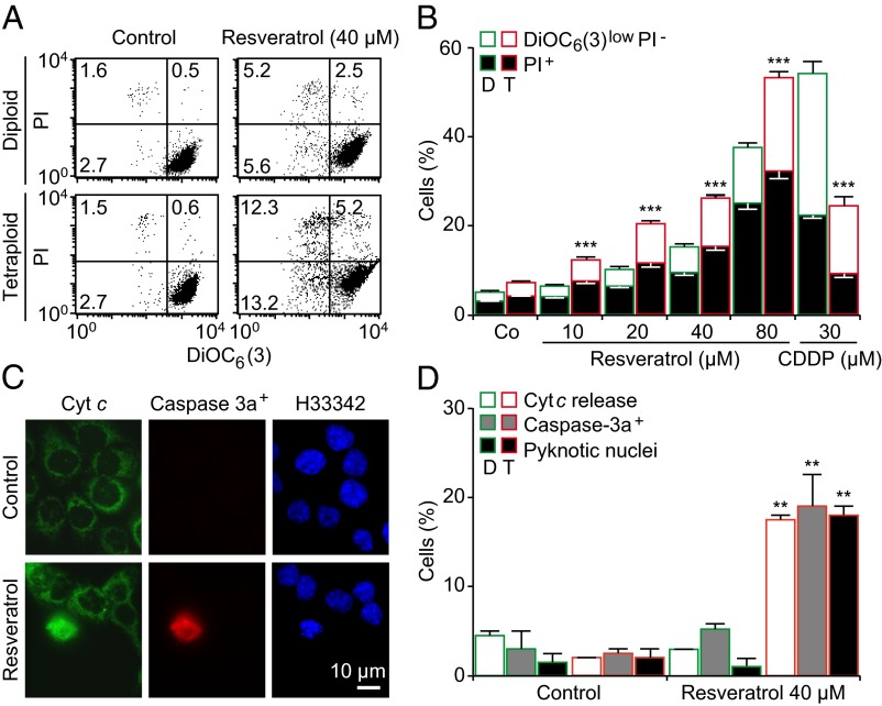
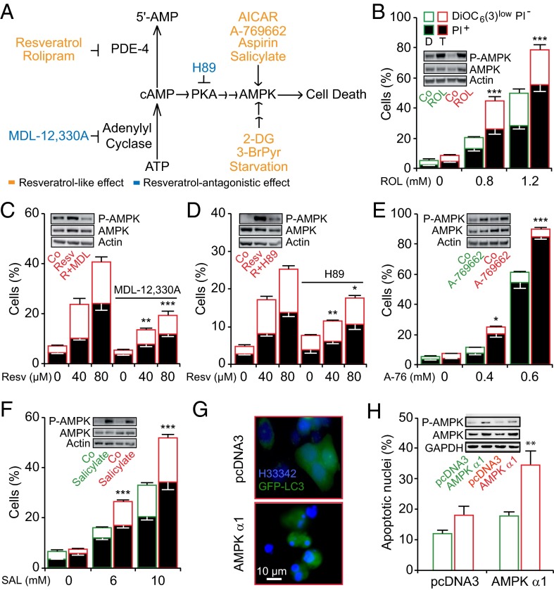
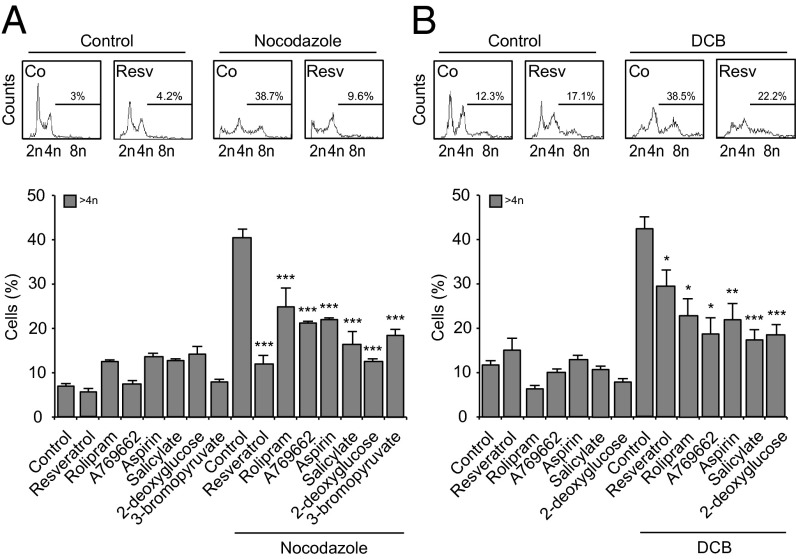
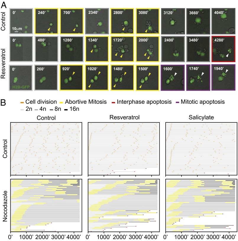
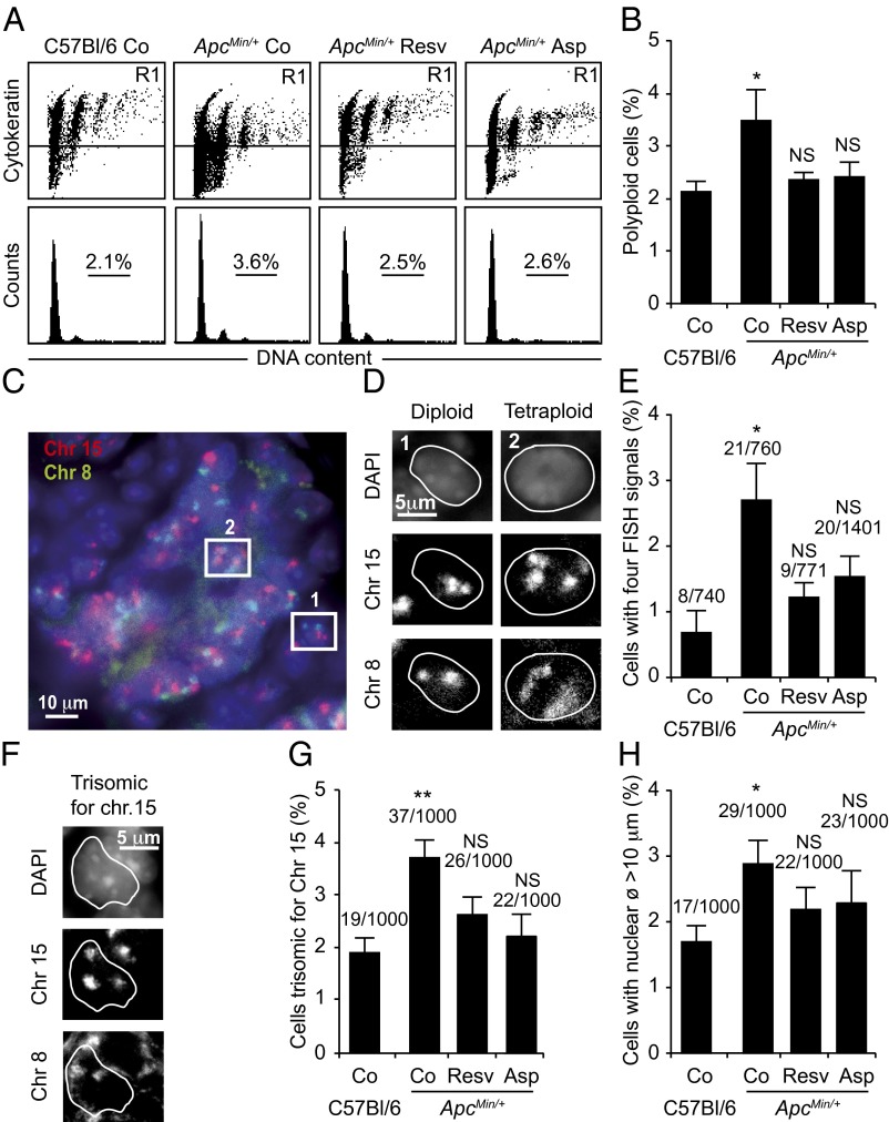
Tetraploidy constitutes a genomically metastable state that can lead to aneuploidy and genomic instability. Tetraploid cells are frequently found in preneoplastic lesions, including intestinal cancers arising due to the inactivation of the tumor suppressor adenomatous polyposis coli (APC). Using a phenotypic screen, we identified resveratrol as an agent that selectively reduces the fitness of tetraploid cells by slowing down their cell cycle progression and by stimulating the intrinsic pathway of apoptosis. Selective killing of tetraploid cells was observed for a series of additional agents that indirectly or directly stimulate AMP-activated protein kinase (AMPK) including salicylate, whose chemopreventive action has been established by epidemiological studies and clinical trials. Both resveratrol and salicylate reduced the formation of tetraploid or higher-order polyploid cells resulting from the culture of human colon carcinoma cell lines or primary mouse epithelial cells lacking tumor protein p53 (TP53, best known as p53) in the presence of antimitotic agents, as determined by cytofluorometric and videomicroscopic assays. Moreover, oral treatment with either resveratrol or aspirin, the prodrug of salicylate, repressed the accumulation of tetraploid intestinal epithelial cells in the Apc(Min/+) mouse model of colon cancer. Collectively, our results suggest that the chemopreventive action of resveratrol and aspirin involves the elimination of tetraploid cancer cell precursors.


Conflict of interest statement

The authors declare no conflict of interest.

Figures

Fig. 1.
Fig. 1.
Validation of the selective inhibitory effect of resveratrol toward tetraploid cells. (AD) Multiple diploid and tetraploid clones from human colon carcinoma HCT116 cells (framed in green and red, respectively) were treated with the indicated concentrations of resveratrol for 48 h before the evaluation of the cell-death–associated parameters either by cytofluorometry upon costaining with the vital dye iodure propidium (PI) and the mitochondrial membrane potential (Δψm)-sensing dye DiOC6(3) (A and B) or by fluorescence microcopy upon coimmunostaining with antibodies directed against cytochrome c (Cyt c) and activated caspase 3 (Caspase-3a) (C and D). A illustrates representative dot plots (numbers refer to the percentage of cells found in each quadrants), whereas B shows quantitative data (mean ± SEM; n = 5) from experiments performed on eight different diploid and seven tetraploid clones. Representative pictures and the quantification of cells displaying Cyt c release, caspase-3 activation (Caspase-3a+), and pyknotic nuclei (as determined by nuclear counterstaining with Hoechst 33342) are reported in C and D, respectively (mean ± SEM; n = 500 cells). In B, the fraction of dying (DiOC6(3)low PI) and dead cells (PI+) is represented by white and black columns, respectively, and cisplatin (CDDP) was used as a negative control. **P < 0.01; ***P < 0.001 (Student t test), compared with the equally treated diploid cells. See also Figs. S2 and S3.
Fig. 2.
Fig. 2.
Activation of the energy sensor 5′AMP-activated protein kinase (AMPK) preferentially induces tetraploid cell death. (A) Proposed model of how resveratrol activates AMPK and induces tetraploid cell death (see text for more details). (BF) Diploid and tetraploid human colon carcinoma HCT116 cells (depicted in green and red, respectively), treated for 48 h with the indicated concentrations of the phosphodiesterase 4 (PDE-4) inhibitor rolipram (B), resveratrol in combination with either the adenylate cyclase inhibitor MDL-12330A (C), or the PKA inhibitor H89 (D), the AMPK activator A-769662 (E), or salicylate (SAL) (F). Thereafter, the cells were subjected to the flow cytometry-assisted measurement of cell death parameters after DiOC6(3)/PI costaining (mean ± SEM; three independent experiments conducted with three diploid and three tetraploid clones). Alternatively, cells were transfected with GFP-LC3 plus vector (pcDNA3) or a cDNA encoding AMPK α1, followed by the quantitation of apoptotic nuclei in transfected (GFP+) cells (G and H). *P < 0.05; **P < 0.01; ***P < 0.001 (Student t test), compared with the equally treated diploid cells (B, E, F, and H) or to cells treated only with resveratrol (C and D). In BF and H, representative immunoblots of phosphorylated AMPK (Thr172) and total AMPK for each drug are depicted as Insets. Actin or GAPDH were used as loading controls. See also Figs. S6 and S8.
Fig. 3.
Fig. 3.
Resveratrol and AMPK activator effects during polyploidization. (A and B) p53-deficient human colon carcinoma HCT116 cells (A) and p53−/− mouse mammary gland epithelial cells (MMECs) (B) were left untreated (control) or incubated for 48 h with the indicated drugs in combination or not with the polyploidizing agents nocodazole 100 nM (A) or dihydrocytochalasin B 1 μM (B). The following concentrations were used (for HCT116 and MMECs, respectively, if different): resveratrol (40 μM), rolipram (0.8 and 1 mM), A-769662 (0.4 and 0.2 mM), aspirin (10 mM), salicylate (10 mM), 2-deoxyglucose (40 and 1 mM), and 3-bromopyruvate (80 μM). At the end of the incubation, DNA content was assessed by cytofluorometry upon Hoechst 33342 staining. Representative cell cycle profiles (Upper) and quantification of polyploid (>4n) cells (Lower) are reported. (Upper) Numbers refer to percentage of polyploid cells. The results are reported as mean ± SEM (n = 3). *P < 0.05; **P < 0.01; ***P < 0.001 (Student t test), compared with the cells exposed to nocodazole or dihydrocytochalasin B alone (Lower). See also Fig. S9.
Fig. 4.
Fig. 4.
Cell fate profiling of newly generated polyploid cells treated with resveratrol or salicylate. (A and B) p53-deficient human colon carcinoma HCT116 cells stably transfected with H2B-GFP were exposed to the indicated drugs combined or not with nocodazole to induce polyploidization. Cell fate was monitored by videomicroscopy for 72 h. Representative pictures of cells treated with nocodazole alone or in combination with resveratrol are shown in A (arrowheads indicate relevant events). In B, the cell fate profiles of 50 cells left untreated (control) or incubated with resveratrol or salicylate, in combination or not with nocodazole are reported. Light gray depicts interphase cell; orange, successful division; yellow, abortive mitosis; red, interphase apoptosis; and purple, mitotic apoptosis. Gray darkening depicts a change in nuclear size and ploidy, from diploidy to decahexaploidy.
Fig. 5.
Fig. 5.
Resveratrol decreases tetraploidy incidence in the intestine of ApcMin/+ mice. (A and B) Cell cycle analyses of cell suspensions. Six-week-old WT C57BL/6 and ApcMin/+ mice were left untreated (Co) or treated with resveratrol (Resv, 100 mg/kg) or aspirin (Asp, 25 mg/kg) for 5 wk. DNA content of cytokeratin+ intestinal crypt cells were analyzed by flow cytometry. Representative dot plots (y axis, cytokeratin; x axis, DNA content) and cell cycle profiles (y axis, cell count; x axis, DNA content) are reported in A, whereas quantification is shown in B for cytokeratin+ cells (mean ± SEM; 10 mice per group). (CH) In situ analyses of paraffin-embedded tissues sections. Intestinal crypt cells then were assessed for their chromosome number by FISH using probes against chromosomes 8 (green) and 15 (orange). A picture of a representative cross-section from an intestinal crypt is shown in C. In D, the areas indicated by two boxes in C are magnified to provide examples of the characteristic pattern of a diploid (Left, 1) and tetraploid (Right, 2) cell. The frequency of tetraploid cells is reported in E. Representative pictures and quantification of trisomic cells for chromosome 15 are shown in F and G, respectively. Cells with a nuclear diameter >10 μm, characteristic of tetraploid cells (Table S1), are reported in H. In E, G, and H, numbers refer to the incidence of the indicated cells per total cells analyzed (mean ± SEM; 5 mice per group). NS, not significant; *P < 0.05, **P < 0.01 (Student t test), compared with WT C57BL/6 controls.

Comment in

References

    1. Ganem NJ, Pellman D. Limiting the proliferation of polyploid cells. Cell. 2007;131(3):437–440. - PubMed
    1. Gordon DJ, Resio B, Pellman D. Causes and consequences of aneuploidy in cancer. Nat Rev Genet. 2012;13(3):189–203. - PubMed
    1. Storchova Z, Kuffer C. The consequences of tetraploidy and aneuploidy. J Cell Sci. 2008;121(Pt 23):3859–3866. - PubMed
    1. Vitale I, et al. Illicit survival of cancer cells during polyploidization and depolyploidization. Cell Death Differ. 2011;18(9):1403–1413. - PMC - PubMed
    1. Vitale I, Galluzzi L, Castedo M, Kroemer G. Mitotic catastrophe: A mechanism for avoiding genomic instability. Nat Rev Mol Cell Biol. 2011;12(6):385–392. - PubMed

Publication types