. 2014 Feb 25;111(8):2978-83.
doi: 10.1073/pnas.1315883111. Epub 2014 Feb 10.

Molecular profiling of Mycobacterium tuberculosis identifies tuberculosinyl nucleoside products of the virulence-associated enzyme Rv3378c

Affiliations

Molecular profiling of Mycobacterium tuberculosis identifies tuberculosinyl nucleoside products of the virulence-associated enzyme Rv3378c

Emilie Layre et al. Proc Natl Acad Sci U S A. .

Abstract

To identify lipids with roles in tuberculosis disease, we systematically compared the lipid content of virulent Mycobacterium tuberculosis with the attenuated vaccine strain Mycobacterium bovis bacillus Calmette-Guérin. Comparative lipidomics analysis identified more than 1,000 molecular differences, including a previously unknown, Mycobacterium tuberculosis-specific lipid that is composed of a diterpene unit linked to adenosine. We established the complete structure of the natural product as 1-tuberculosinyladenosine (1-TbAd) using mass spectrometry and NMR spectroscopy. A screen for 1-TbAd mutants, complementation studies, and gene transfer identified Rv3378c as necessary for 1-TbAd biosynthesis. Whereas Rv3378c was previously thought to function as a phosphatase, these studies establish its role as a tuberculosinyl transferase and suggest a revised biosynthetic pathway for the sequential action of Rv3377c-Rv3378c. In agreement with this model, recombinant Rv3378c protein produced 1-TbAd, and its crystal structure revealed a cis-prenyl transferase fold with hydrophobic residues for isoprenoid binding and a second binding pocket suitable for the nucleoside substrate. The dual-substrate pocket distinguishes Rv3378c from classical cis-prenyl transferases, providing a unique model for the prenylation of diverse metabolites. Terpene nucleosides are rare in nature, and 1-TbAd is known only in Mycobacterium tuberculosis. Thus, this intersection of nucleoside and terpene pathways likely arose late in the evolution of the Mycobacterium tuberculosis complex; 1-TbAd serves as an abundant chemical marker of Mycobacterium tuberculosis, and the extracellular export of this amphipathic molecule likely accounts for the known virulence-promoting effects of the Rv3378c enzyme.

Keywords: TbAd; terpenyl transferase.


Conflict of interest statement

The authors declare no conflict of interest.

Figures
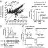
Fig. 1.
Fig. 1.
Comparative lipidomic analysis of M. tuberculosis and BCG reveals a natural product constitutively produced and exported by M. tuberculosis. (A) Detected molecular features are shown as a scatterplot of intensity derived from M. tuberculosis H37Rv and BCG lipid extracts. Each feature corresponds to a detected ion and contains retention time and m/z values, which are detailed in SI Appendix, Dataset S1; 1,845 features out of 7,852 total features showed intensity ratios that deviate significantly from 1 (corrected P value, <0.05). The mass spectrum corresponds to the four M. tuberculosis-specific features of substance A. (B) Ion chromatograms extracted at m/z (540.3545) and retention time of substance A were used for the analysis of lipid extracts of reference strains. (C) Ion chromatograms from lipidomic analysis of filtered conditioned medium were extracted at the m/z of substance A or control compounds that are secreted (carboxymycobactin) and cell wall-associated lipids (trehalose monomycolate, mycobactin).
Fig. 2.
Fig. 2.
Identification of 1-TbAd. The structure of substance A purified from M. tuberculosis lipid extract was characterized using CID-MS and NMR (800 MHz) analyses yielding key collision products and resonances detailed in SI Appendix, Figs. S2–S9.
Fig. 3.
Fig. 3.
M. tuberculosis biosynthesis of substance A requires Rv3378c. (A) The screening of 4,196 transposon mutants of M. tuberculosis H37Rv using a rapid 3-min HPLC-MS method yielded 30 strains with reduced 1-TbAd signal. (B) Rescreening with the 40-min HPLC-MS method confirmed absence of 1-TbAd signal in two mutants. (C) Both mutants were found to have spontaneous, non–transposon-induced mutations in Rv3378c and were subject to complementation of Rv3377c-Rv3378c and reanalysis for 1-TbAd production.
Fig. 4.
Fig. 4.
Rv3378c acts as a tuberculosinyl transferase. (A) Rv3377c and Rv3378c are currently thought to produce tuberculosinol and isotuberculosinol. (B) The existence of 1-TbAd might be explained by a revised function of the Rv3378c enzyme, which acts as a tuberculosinyl transferase. (C and D) Ion chromatograms and mass spectra (Insets) (C) and CID-MS (D) of the 1-TbAd standard and reaction products of enzymatic assays performed using recombinant Rv3378c protein.
Fig. 5.
Fig. 5.
The expression of Rv3377c-Rv3378c is sufficient for production of 1-TbAd in M. smegmatis. Extracted ion chromatograms and mass spectra (Insets) of 1-TbAd (m/z 540.3545) for the HPLC-MS analysis of lipid extracts from M. tuberculosis (Left), M. smegmatis parental (Center), or each of three M. smegmatis Rv3377c-Rv3378c knock-in (Right) strains.
Fig. 6.
Fig. 6.
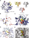
Rv3378c adopts a (Z)-prenyl transferase fold. (A) Structure of the Rv3378c dimer is compared with conventional (Z)-prenyl transferases. (B) Superposition of the active site of Rv3378c and other (Z)-prenyl transferases with the pyrophosphate bound to Rv2361c (stick) shows conserved key residues for substrate binding and catalysis (Rv3378c, blue; Rv2361c, yellow; Rv1086, gray; E. coli UPP synthase, magenta for carbon atoms). (C) The monomeric subunits of Rv3378c and Rv2361c were superimposed and Rv2361c substrates (sphere) (carbon, yellow/gray; oxygen, red; phosphate, orange) are modeled in the active site of Rv3378c. The conserved residue, Asp34, is shown as a stick model, and the magnesium ion is shown as a magenta sphere. (D) Proposed model of Rv3378c shows two substrate pockets with hydrophobic residues lining the predicted prenyl binding pocket and D34 positioned adjacent to the predicted adenosine binding pocket. (BD) The flexible P-loop of Rv3378c (residues 80–95) is colored in red, with the dotted line for disordered region (residues 84–90). (E) The translucent surface of Rv3378c was modeled with substrates (spheres) using the same view as D.

References

    1. Dye C, Glaziou P, Floyd K, Raviglione M. Prospects for tuberculosis elimination. Annu Rev Public Health. 2013;34:271–286. - PubMed
    1. Sturgill-Koszycki S, et al. Lack of acidification in Mycobacterium phagosomes produced by exclusion of the vesicular proton-ATPase. Science. 1994;263(5147):678–681. - PubMed
    1. Camus JC, Pryor MJ, Médigue C, Cole ST. Re-annotation of the genome sequence of Mycobacterium tuberculosis H37Rv. Microbiology. 2002;148(Pt 10):2967–2973. - PubMed
    1. Layre E, et al. A comparative lipidomics platform for chemotaxonomic analysis of Mycobacterium tuberculosis. Chem Biol. 2011;18(12):1537–1549. - PMC - PubMed
    1. Sartain MJ, Dick DL, Rithner CD, Crick DC, Belisle JT. Lipidomic analyses of Mycobacterium tuberculosis based on accurate mass measurements and the novel “Mtb LipidDB”. J Lipid Res. 2011;52(5):861–872. - PMC - PubMed

Publication types

MeSH terms

LinkOut - more resources