

Sparganosis presenting as cauda equina syndrome with molecular identification of the parasite in tissue sections
- PMID: 24516282
- PMCID: PMC3916466
- DOI: 10.3347/kjp.2013.51.6.739
Sparganosis presenting as cauda equina syndrome with molecular identification of the parasite in tissue sections
Abstract
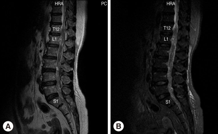
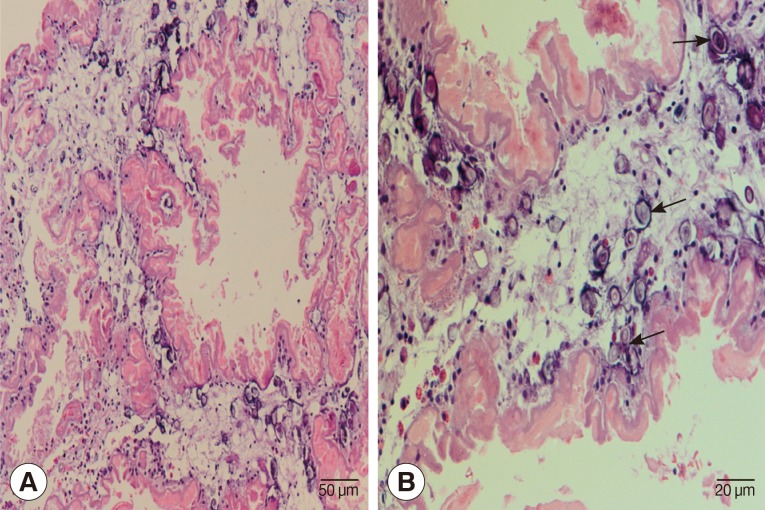
A 52-year-old woman presented with lower back pain, progressive symmetrical paraparesis with sensory impairment, and sphincter disturbance. Magnetic resonance imaging (MRI) of the whole spine revealed multiple intradural extramedullary serpiginous-mass lesions in the subarachnoid space continuously from the prepontine to the anterior part of the medulla oblongata levels, C7, T2-T8, and T12 vertebral levels distally until the end of the theca sac and filling-in the right S1 neural foramen. Sparganosis was diagnosed by demonstration of the sparganum in histopathological sections of surgically resected tissues and also by the presence of serum IgG antibodies by ELISA. DNA was extracted from unstained tissue sections, and a partial fragment of mitochondrial cytochrome c oxidase subunit 1 (cox1) gene was amplified using a primer set specific for Spirometra spp. cox1. After sequencing of the PCR-amplicon and alignment of the nucleotide sequence data, the causative agent was identified as the larva of Spirometra erinaceieuropaei.
Keywords: Spirometra erinaceieuropaei; cauda equina syndrome; molecular identification; sparganosis.
Figures
References
-
- Waeschenbach A, Webster BL, Bray RA, Littlewood DT. Added resolution among ordinal level relationships of tapeworms (Platyhelminthes: Cestoda) with complete small and large subunit nuclear ribosomal RNA genes. Mol Phylogenet Evol. 2007;45:311–325. - PubMed
-
- Kuchta R, Scholz T, Bray RA. Revision of the order Bothriocephalidea Kuchta, Scholz, Brabec & Bray, 2008 (Eucestoda) with amended generic diagnoses and keys to families and genera. Syst Parasitol. 2008;71:81–136. - PubMed
-
- Beaver PC, Jung RC, Cupp EW. Clinical Parasitology. 9th ed. Philadelphia, USA: Lea & Febiger; 1984. pp. 494–504.
-
- Miyazaki I. An illustrated book of Helminthic Zoonoses. Tokyo, Japan: International Medical Foundation of Japan; 1991. pp. 207–214.
-
- Chang KH, Han MH. MRI of CNS parasitic diseases. J Magn Reson Imaging. 1998;8:297–307. - PubMed
Publication types
MeSH terms
Substances
Associated data
- Actions
LinkOut - more resources
Full Text Sources
Other Literature Sources
Miscellaneous
