. 2014 Feb 6;10(2):e1003991.
doi: 10.1371/journal.pgen.1003991. eCollection 2014 Feb.

Canine hereditary ataxia in old english sheepdogs and gordon setters is associated with a defect in the autophagy gene encoding RAB24

Affiliations

Canine hereditary ataxia in old english sheepdogs and gordon setters is associated with a defect in the autophagy gene encoding RAB24

Caryline Agler et al. PLoS Genet. .

Abstract

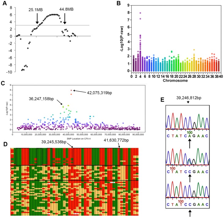
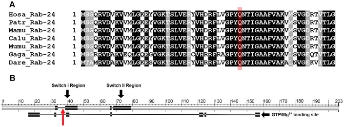
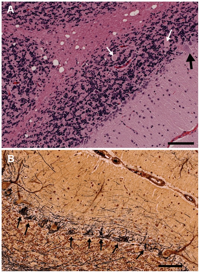
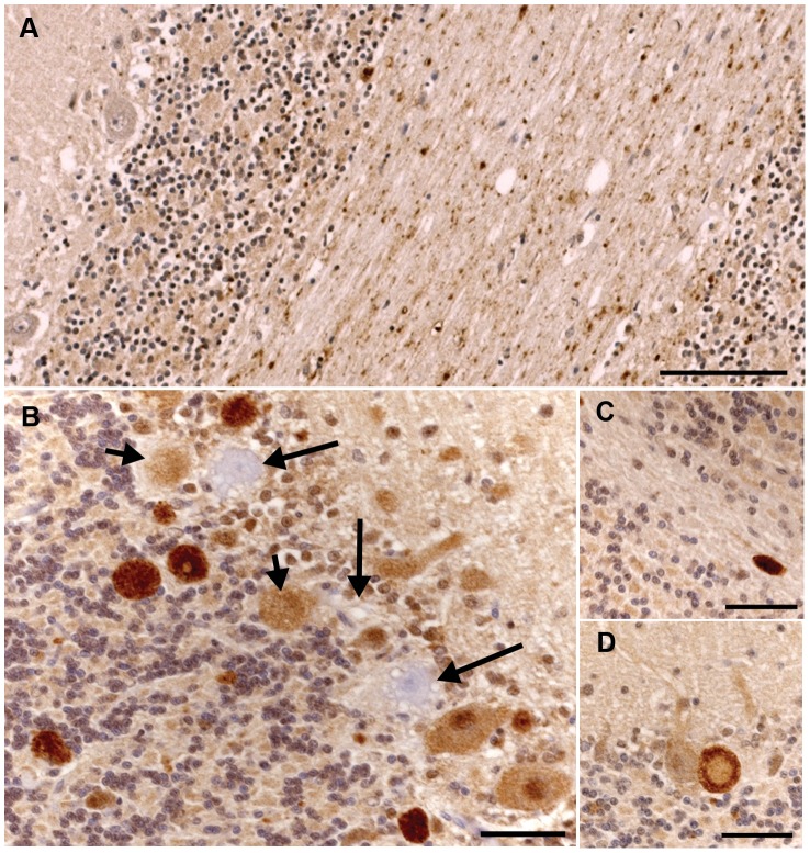
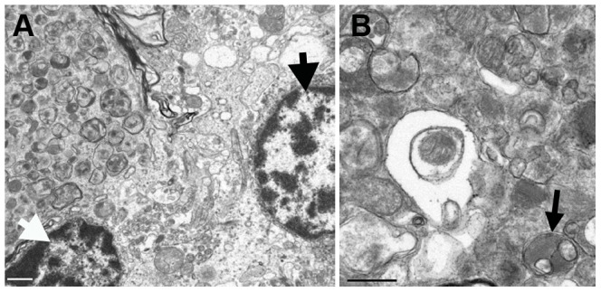
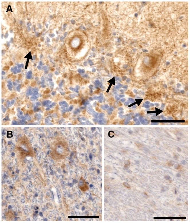
Old English Sheepdogs and Gordon Setters suffer from a juvenile onset, autosomal recessive form of canine hereditary ataxia primarily affecting the Purkinje neuron of the cerebellar cortex. The clinical and histological characteristics are analogous to hereditary ataxias in humans. Linkage and genome-wide association studies on a cohort of related Old English Sheepdogs identified a region on CFA4 strongly associated with the disease phenotype. Targeted sequence capture and next generation sequencing of the region identified an A to C single nucleotide polymorphism (SNP) located at position 113 in exon 1 of an autophagy gene, RAB24, that segregated with the phenotype. Genotyping of six additional breeds of dogs affected with hereditary ataxia identified the same polymorphism in affected Gordon Setters that segregated perfectly with phenotype. The other breeds tested did not have the polymorphism. Genome-wide SNP genotyping of Gordon Setters identified a 1.9 MB region with an identical haplotype to affected Old English Sheepdogs. Histopathology, immunohistochemistry and ultrastructural evaluation of the brains of affected dogs from both breeds identified dramatic Purkinje neuron loss with axonal spheroids, accumulation of autophagosomes, ubiquitin positive inclusions and a diffuse increase in cytoplasmic neuronal ubiquitin staining. These findings recapitulate the changes reported in mice with induced neuron-specific autophagy defects. Taken together, our results suggest that a defect in RAB24, a gene associated with autophagy, is highly associated with and may contribute to canine hereditary ataxia in Old English Sheepdogs and Gordon Setters. This finding suggests that detailed investigation of autophagy pathways should be undertaken in human hereditary ataxia.


Conflict of interest statement

I have read the journal's policy and have the following conflicts. The corresponding author's (Dr. Olby's) laboratory is offering a genetic test for this disease in Gordon Setters and Old English Sheepdog. The proceeds do not return to the author but are used to support ongoing research. The test has not been and will not be patented.

Figures

Figure 1
Figure 1. Pedigree of family of Old English Sheepdogs genotyped with microsatellite markers for linkage analysis.
Red: genotyped. Solid symbol: cases, open symbol: controls, hashed symbol: unknown.
Figure 2
Figure 2. Results of genotyping, linkage and genome-wide association analyses.
All coordinates refer to CanFam 2. a) LOD scores on CFA 4. LOD scores >3 were found between markers 408 and REN74B1 located at 25.1 and 44.8 MB respectively. b) A Manhattan plot of −log10 p-value against chromosome. This genome-wide association study using 12,986 SNPs showed significant association on CFA4. c) On CFA4 SNPs with significant association after Bonferroni correction were located between 36 and 42 MB. d) The SNP genotypes are shown in a chart. Each row is a different individual and each column is a different SNP. Solid green and red boxes indicate homozygosity while yellow boxes indicate heterozygosity. The genotype of cases is displayed in the top segment of the chart, their parents are shown in the middle section between the two solid, horizontal black lines, and the remaining phenotypically normal dogs in the bottom segment of the chart. A large region of homozygosity extends from 35,402,483 bp to 41,772,116 bp in all affected dogs and smaller regions in which the affected dogs are identical and different from the normal dogs are indicated by black boxes. * lies over the two most significant SNPs from the GWAS. e) Sequence of a portion of RAB24 showing the A>C mutation. Arrows under the traces indicate the target nucleotide. Normal wild type dogs are shown in the top electropherogram, heterozygotes in the middle electropherogram and homozygotes for the mutation in the bottom electropherogram.
Figure 3
Figure 3. The Rab24 protein and mutation location.
a) ClustalW multiple protein alignment of the region of Rab24 containing the mutation. The target glutamine is highlighted in red. Sequences for humans (Hosa), chimpanzees (Patr), rhesus macaque (Mamu), wolf (Calu), mouse (Mumu), chicken (Gaga) and zebrafish (Dare) are shown. A black background indicates 100% homology in these species; the target glutamine lies within a region of 14 highly conserved amino acids. b) Proposed switch I and II regions are shown superimposed on the Rab24 protein and the GTPase/Mg2+ binding site is shown below the protein. The mutation at the 38th amino acid lies within switch I region and the GTPase/Mg2+ binding site (red arrow).
Figure 4
Figure 4. Sections of the cerebellum from an affected 2.5-year-old Gordon Setter stained with hematoxylin and eosin (a) and Bielschowsky (b).
a) The three cortical layers and cerebellar cortical white matter are shown. There is dramatic loss of Purkinje neurons with only one Purkinje neuron (black arrow) visible at the junction of the granular and molecular layers. Axonal spheroids can be seen within the granular layer (white arrows) and there is marked vacuolation of the white matter. Bar = 100 µm. b) The Bielschowsky stain highlights the processes of basket cells around Purkinje neurons, thus emphasizing the loss of Purkinje neurons (black arrows).
Figure 5
Figure 5. Ubiquitin immunohistochemical staining of the cerebellum of an unaffected 11-year-old Dachshund and an affected 2.5-year-old Gordon Setter.
a) There is granular ubiquitin positive material in the white matter tracts of the cerebellum in this unaffected dog. The Purkinje neurons are negative. Bar = 100 µm. b) There are multiple large, intensely ubiquitin positive circular bodies in the granular layer and at the junction of the molecular and granular layers in this affected Gordon Setter. The ubiquitin staining has a punctate character within these bodies. In addition, there are Purkinje and molecular neurons with diffuse cytoplasmic and nuclear ubiquitin staining. Note also the three highly vacuolated Purkinje neurons that are negative for ubiquitin (long arrows). Two of these degenerating Purkinje neurons have ubiquitin positive circular bodies immediately adjacent to them (short arrows). These circular bodies correlated to axonal spheroids on the hematoxylin and eosin stains and, as shown in figure 5d, appeared to connect directly to Purkinje neurons in some instances. Bar = 50 µm. c) An example of a large ovoid ubiquitin positive body in the cerebellar white matter of the 2.5-year-old affected dog. Bar = 50 µm. d) A ubiquitin positive body lies immediately adjacent to (and apparently connecting with) a Purkinje neuron and has a less intensely staining center. Bar = 50 µm.
Figure 6
Figure 6. Transmission electron micrographs of the cerebellum of an affected Gordon Setter.
a) A partially-captured axonal spheroid, located at the junction of the Purkinje and granular layers of the cerebellum, packed with late autophagosomes, is visible in the upper left hand side of the image and it is closely associated with a Purkinje neuron (thick black arrow) and a granular neuron (thick white arrow). Bar = 1 µm. b) At higher magnification, the wide array of morphologies of autophagosomes can be seen along with some more normal appearing mitochondria. Autophagosomes typically have a double membrane and an example of a degradative autophagosome is illustrated here (black arrow). Bar = 0.5 µm.
Figure 7
Figure 7. Rab24 immunohistochemical staining of the cerebellum of an affected dog.
a) There is intense eccentric perinuclear staining of the Purkinje cells and the processes of the basket cells. The basket cell staining can still be seen in places where the Purkinje neurons have died (arrows). b) The neurons in the deep cerebellar nuclei all exhibit cytoplasmic staining for Rab24. c) Cells stained positive within the white matter of the cerebellum. Due to their morphology, these cells are believed to be oligodendrocytes. Bar = 100 µm in all images.

References

    1. Klockgether T (2011) Paulson H (2011) Milestones in ataxia. Mov Disord 26: 1134–1141. - PMC - PubMed
    1. Klockgether T (2012) Sporadic adult-onset ataxia of unknown etiology. Handb Clin Neurol 103: 253–262. - PubMed
    1. Hersheson J, Haworth A (2012) Houlden H (2012) The inherited ataxias: Genetic heterogeneity, mutation databases, and future directions in research and clinical diagnostics. Hum Mutat 33: 1324–1332. - PubMed
    1. Bird TD (2013) Hereditary Ataxia Overview. In: Pagon RA, Bird TD, Dolan CR, Stephens K, Adam MP, editors. GeneReviews. Seattle: University of Washington. - PubMed
    1. Seidel K, Siswanto S, Brunt ER, den Dunnen W, Korf HW, et al. (2012) Brain pathology of spinocerebellar ataxias. Acta Neuropathol 124: 1–21. - PubMed

Substances

LinkOut - more resources