. 2014 Feb 7;9(2):e87577.
doi: 10.1371/journal.pone.0087577. eCollection 2014.

High-resolution structure of the N-terminal endonuclease domain of the Lassa virus L polymerase in complex with magnesium ions

Affiliations

High-resolution structure of the N-terminal endonuclease domain of the Lassa virus L polymerase in complex with magnesium ions

Gregor D Wallat et al. PLoS One. .

Abstract

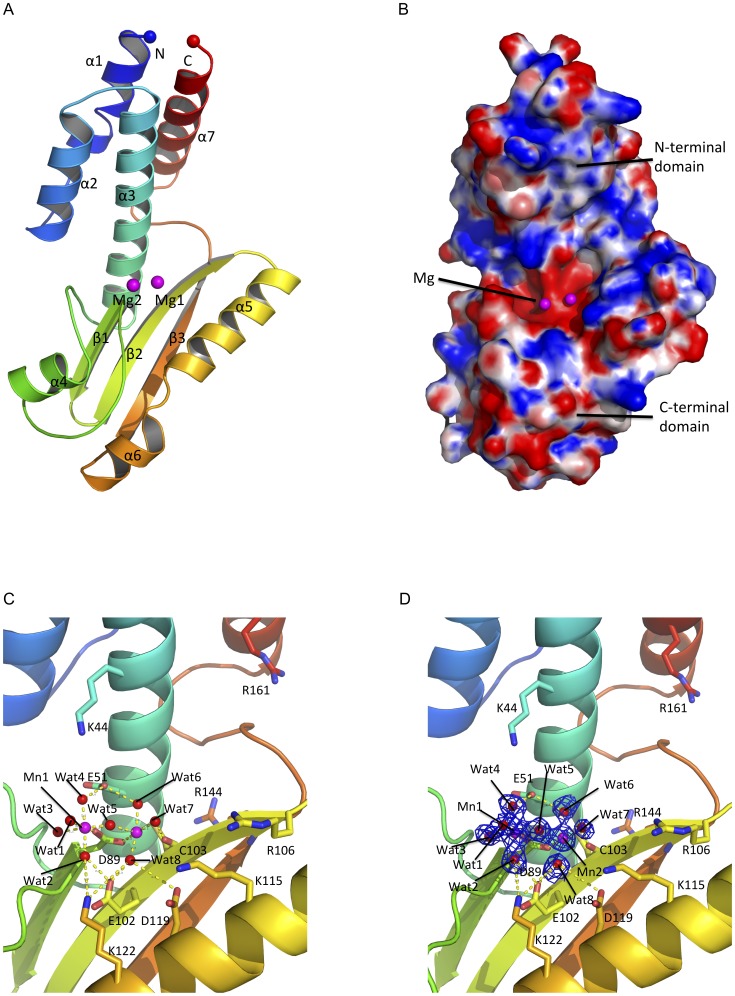
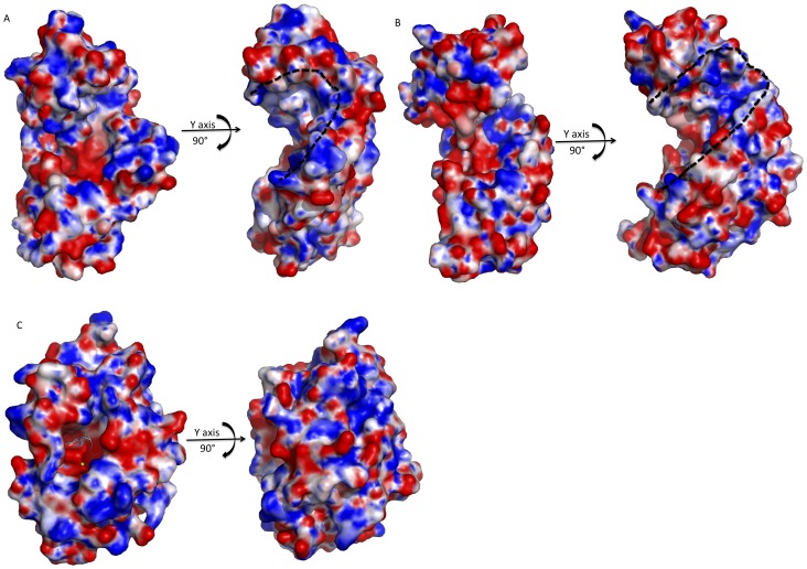
Lassa virus (LASV) causes deadly hemorrhagic fever disease for which there are no vaccines and limited treatments. LASV-encoded L polymerase is required for viral RNA replication and transcription. The functional domains of L-a large protein of 2218 amino acid residues-are largely undefined, except for the centrally located RNA-dependent RNA polymerase (RdRP) motif. Recent structural and functional analyses of the N-terminal region of the L protein from lymphocytic choriomeningitis virus (LCMV), which is in the same Arenaviridae family as LASV, have identified an endonuclease domain that presumably cleaves the cap structures of host mRNAs in order to initiate viral transcription. Here we present a high-resolution crystal structure of the N-terminal 173-aa region of the LASV L protein (LASV L173) in complex with magnesium ions at 1.72 Å. The structure is highly homologous to other known viral endonucleases of arena- (LCMV NL1), orthomyxo- (influenza virus PA), and bunyaviruses (La Crosse virus NL1). Although the catalytic residues (D89, E102 and K122) are highly conserved among the known viral endonucleases, LASV L endonuclease structure shows some notable differences. Our data collected from in vitro endonuclease assays and a reporter-based LASV minigenome transcriptional assay in mammalian cells confirm structural prediction of LASV L173 as an active endonuclease. The high-resolution structure of the LASV L endonuclease domain in complex with magnesium ions should aid the development of antivirals against lethal Lassa hemorrhagic fever.


Conflict of interest statement

Competing Interests: The authors have declared that no competing interests exist.

Figures

Figure 1
Figure 1. Structure of the LASV L endonuclease domain.
A, The cartoon representation of the LASV endonuclease domain is shown in rainbow colors. The N-terminus is shown as a blue sphere and the C-terminus is shown as a red sphere. Magnesium ions are shown as magenta spheres. B, Electrostatic potential map of LASV L endonuclease domain. The highly positive charged residues are shown in blue (+10 KbT/ec) and highly negatively charged residues shown in red (−10KbT/ec). The catalytic cavity is located in vicinity to the magnesium ions. The putative RNA binding cleft is formed diagonally between the N-terminal and the C-terminal domains of the endonuclease. C, Atomic view of the active site of the LASV L N-terminal endonuclease domain. The two magnesium ions are coordinated by water molecules and the side chains of amino acid residue D89 and the main chains of C103. D, The original FoFc electron density map of the two magnesium ions and the water molecules contoured at 3σ.
Figure 2
Figure 2. Similarities and differences between the structures of LASV L endonuclease domain and those of other negative-strand RNA viruses.
Like in figure 1, the LASV L endonuclease domain is rainbow-colored. A, Structure of the LASV L endonuclease overlapping LCMV L endonuclease domain (orange). The residues are numbered based on the LASV L sequence. B, Superimposition of the structure of the LASV L endonuclease domain with the LACV L endonuclease domain (magenta). The magnesium ion of LACV is shown in gray, which is close to the position of the first magnesium ion in the LASV structure. C, Superimposition of LASV L endonuclease domain with influenza PA endonuclease domain (gray). The magnesium ion (gray sphere) from the structure of the influenza PA endonuclease is located close to the second magnesium ion of the LASV L endonuclease.
Figure 3
Figure 3. In vitro endonuclease activity of LASV L173.
A, A 5′ FAM-labeled 16-nt single-stranded RNA substrate was incubated in a buffer with or without purified LASV endonuclease, with or without Mg2+ and with or without metal ion chelator EDTA, for 20 min at 37°C. The reaction products were separated by urea-PAGE and detected by fluorescence scanning. B, The in vitro endonuclease assay was conducted in buffers with different divalent cations for either 5 or 20 min. C, Percentage of RNA substrate degradation after 20 min incubation in a buffer with different divalent cations was quantified by fluorescence scanning. D, WT or mutant LASV L173 was analyzed by an in vitro endonuclease assay for 0, 25, 40 and 90 min. E, Percentage of RNA substrate degradation after 25 min was quantified by fluorescence scanning and normalized to WT control (set at 100%).
Figure 4
Figure 4. Mutational analysis of LASV L endonuclease domain in the LASV minigenomic RNA transcription assay.
FLAG-tagged L expression vector with either WT or alanine substitution at the respective residue within the N-terminal endonuclease domain was used to transfect 293T cells, together with the myc-tagged NP expression vector and LUC-encoded LASV MG RNA. LUC activity was measured and plotted in log scale. Results shown are the average of at least three independent experiments with error bars representing standard deviations. The expression of L (WT or mutant), NP, and GAPDH in the transfected cells was detected by Western blot analysis using anti-FLAG, anti-myc, and anti-GAPDH antibody, respectively.
Figure 5
Figure 5. Electrostatic potential maps of the putative RNA binding cleft of known viral endonucleases.
The black dotted lines outline the potential RNA binding clefts. A, The electrostatic potential maps of LCMV endonuclease. The putative RNA binding cleft were revealed by rotation by 90° along the y axis. B, The electrostatic potential map of LACV endonuclease. Rotation by 90° along the y axis shows the putative RNA binding cleft. C, The electrostatic potential maps of influenza PA endonuclease. The RNA binding cleft of the influenza endonuclease is closed/hidden. Rotation by 90° along the y-axis could not see the putative RNA binding cleft.

References

    1. Khan SH, Goba A, Chu M, Roth C, Healing T, et al. (2008) New opportunities for field research on the pathogenesis and treatment of Lassa fever. Antiviral Res 78: 103–15. - PubMed
    1. McCormick JB, King IJ, Webb PA, Scribner CL, Craven RB, et al. (1986) Lassa fever. Effective therapy with ribavirin. N Engl J Med 314: 20–6. - PubMed
    1. Amorosa V, MacNeil A, McConnell R, Patel A, Dillon KE, et al. (2010) Imported Lassa fever, Pennsylvania, USA, 2010. Emerg Infect Dis 16: 1598–600. - PMC - PubMed
    1. Atkin S, Anaraki S, Gothard P, Walsh A, Brown D, et al. (2009) The first case of Lassa fever imported from Mali to the United Kingdom, February 2009. Euro Surveill 14. - PubMed
    1. Hirabayashi Y, Oka S, Goto H, Shimada K, Kurata T, et al. (1989) The first imported case of Lassa fever in Japan. Nihon Rinsho 47: 71–5. - PubMed

Publication types

MeSH terms