Review
. 2014 Feb 6;9(2):e87737.
doi: 10.1371/journal.pone.0087737. eCollection 2014.

Global diversity and review of Siphonophorae (Cnidaria: Hydrozoa)

Affiliations
Review

Global diversity and review of Siphonophorae (Cnidaria: Hydrozoa)

Gillian M Mapstone. PLoS One. .

Erratum in

Abstract

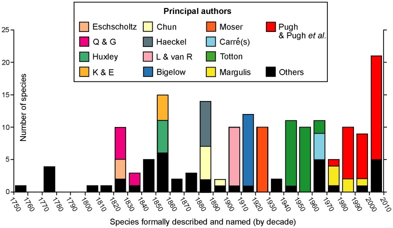
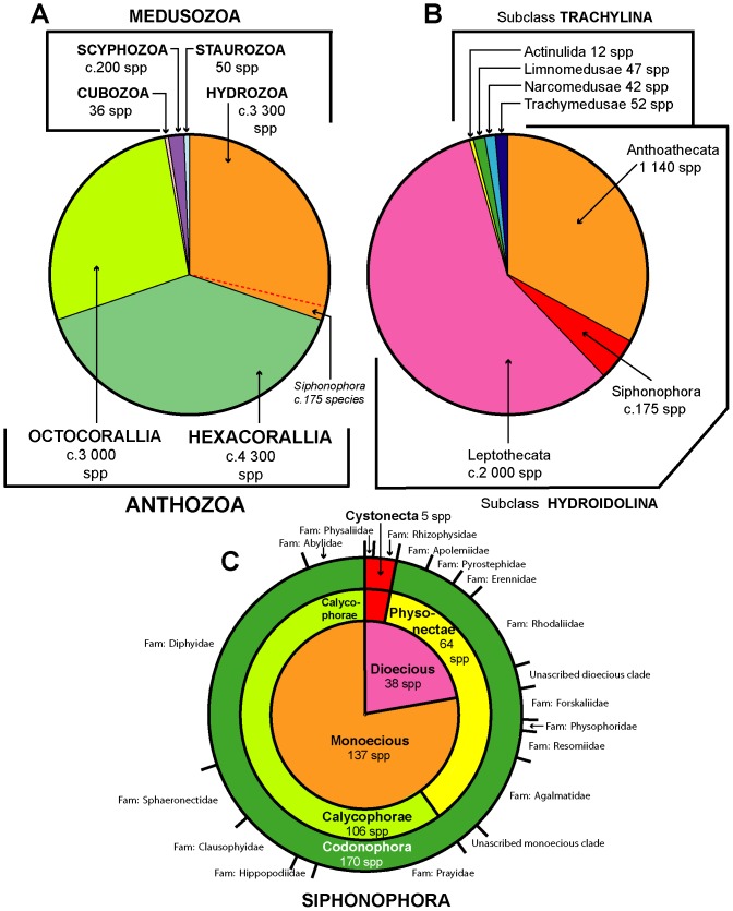
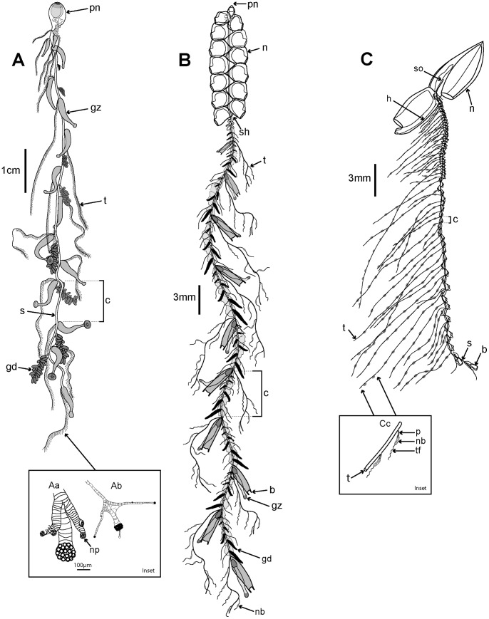
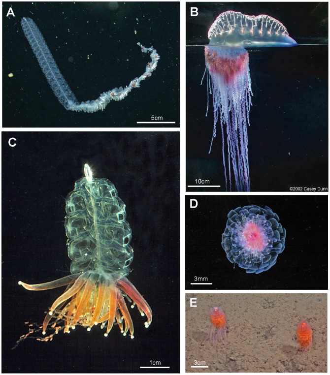
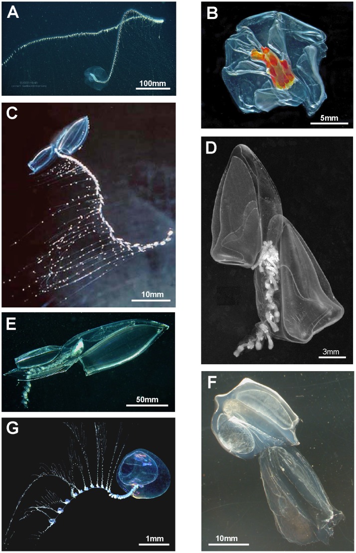
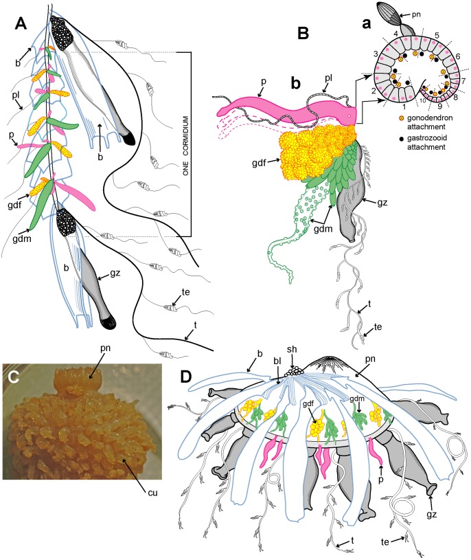
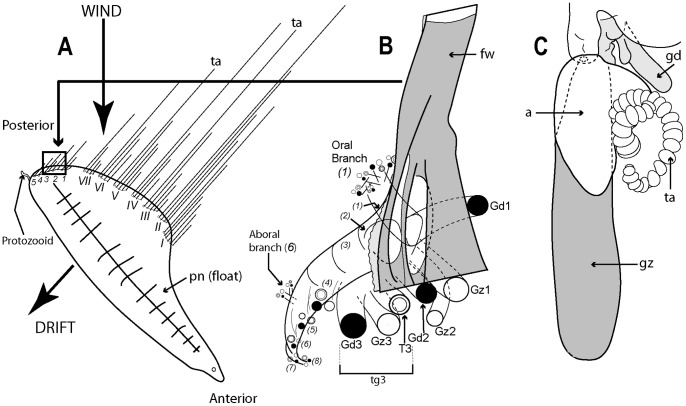
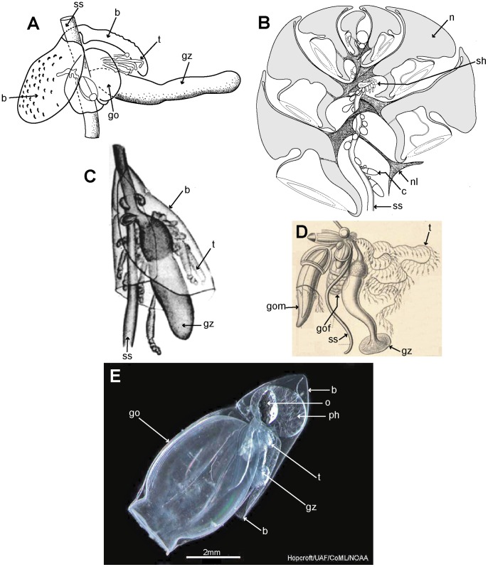
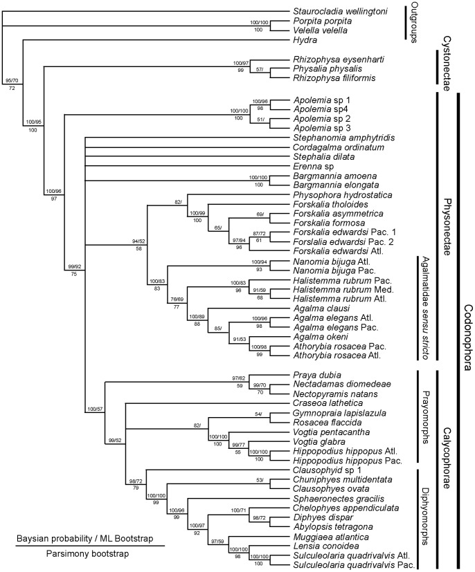
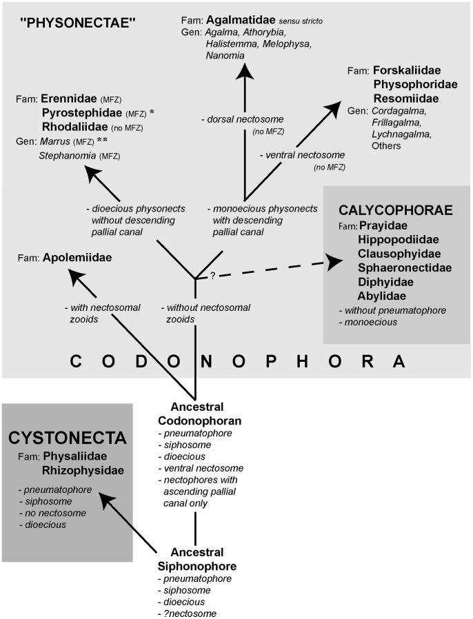
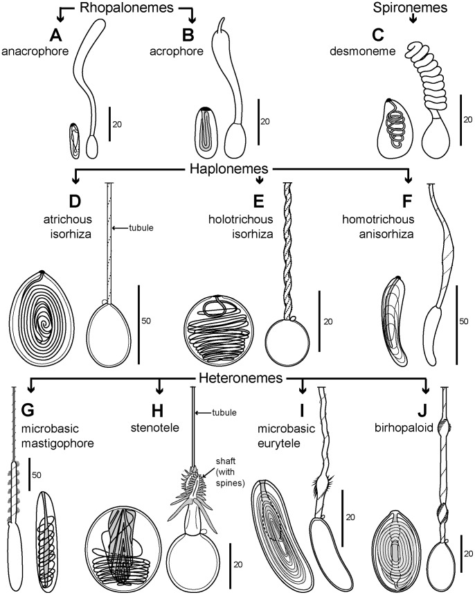
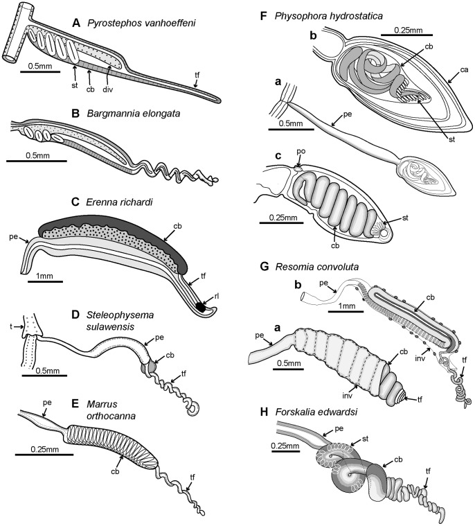
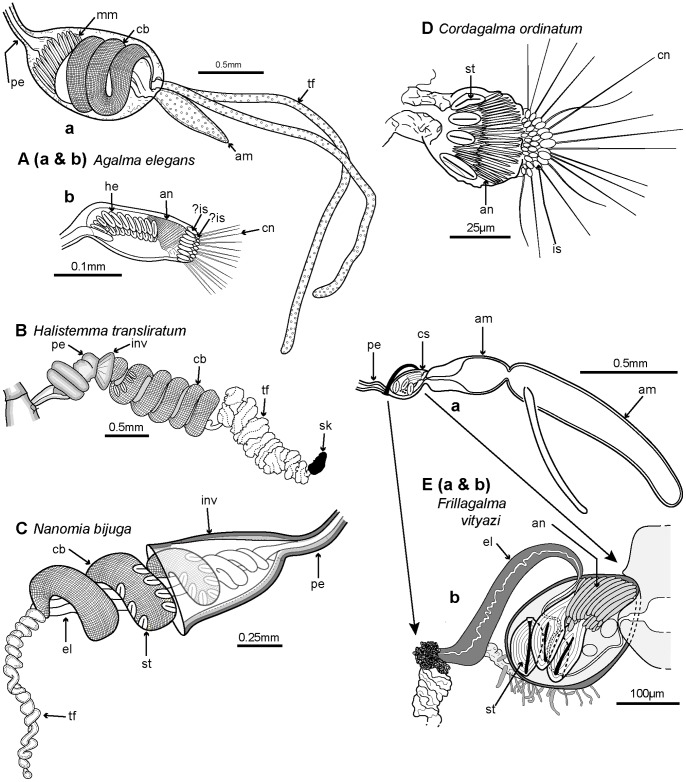
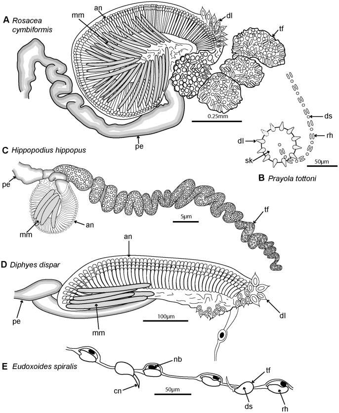
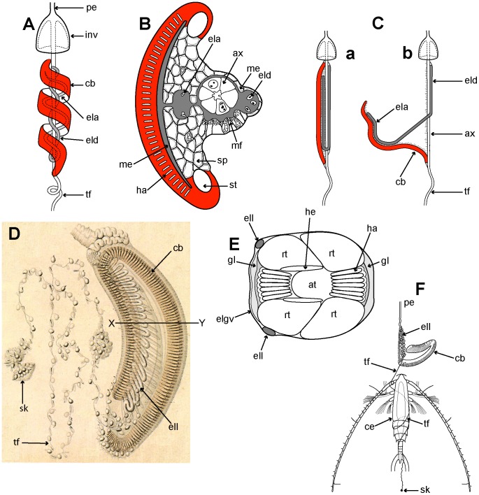
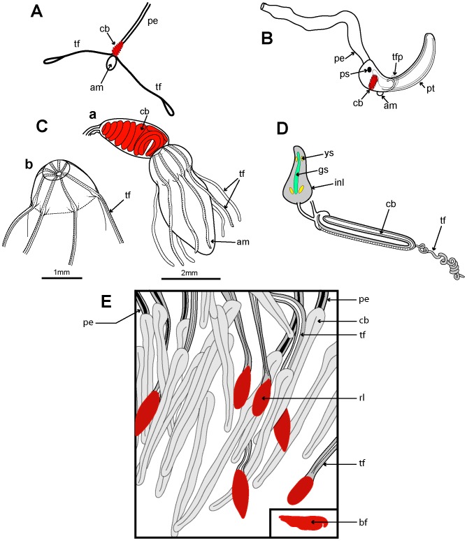
In this review the history of discovery of siphonophores, from the first formal description by Carl Linnaeus in 1785 to the present, is summarized, and species richness together with a summary of world-wide distribution of this pelagic group within the clade Hydrozoa discussed. Siphonophores exhibit three basic body plans which are briefly explained and figured, whilst other atypical body plans are also noted. Currently, 175 valid siphonophore species are recognized in the latest WoRMS world list, including 16 families and 65 genera. Much new information since the last review in 1987 is revealed from the first molecular analysis of the group, enabling identification of some new morphological characters diagnostic for physonect siphonophores. Ten types of nematocysts (stinging cells) are identified in siphonophores, more than in any other cnidarian; these are incorporated into batteries in the side branches of the tentacles in most species (here termed tentilla), and tentilla are reviewed in the last section of this paper. Their discharge mechanisms are explained and also how the tentilla of several physonect siphonophores are modified into lures. Of particular interest is the recent discovery of a previously unknown red fluorescent lure in the tentilla of the deep sea physonect Erenna, the first described example of emission of red light by an invertebrate to attract prey.


Conflict of interest statement

Competing Interests: The author has declared that no competing interests exist.

Figures

Figure 1
Figure 1. History of siphonophore research.
Principle researchers and others from mid-18th century to the present. Authors identified only by initials are Q & G: Quoy and Gaimard, K & E: Keferstein and Ehlers, and L & van R: Lens and van Riemsdijk.
Figure 2
Figure 2. Cnidaria and Siphonophora Species Richness.
A: the c. 11,000 Cnidaria species (excluding Myxozoa) subdivided into clades following Kayal et al. ; B: the c. 3,300 Hydrozoa species, subdivided into ranks from Daly et al. and the present work; C: the 175 valid Siphonophora species subdivided into ranks based on Tables 3 and 4 of the present work.
Figure 3
Figure 3. Three typical siphonophore body plans.
A. Long-stemmed cystonect Rhizophysa eysenhardti (derived from pl. 14 fig. 1): inset shows nematocyst pads on two interpretations of tricornuate tentacular side branches from Rhizophysa filiformis, (Aa: derived from fig. 5 and Ab: derived from pl. 4, fig. 2): B. Long-stemmed physonect Nanomia bijuga (derived from , pl. 7, fig. 1); C. Typical calycophoran Lensia conoidea (derived from photo image by Rob Sherlock - shown in Fig. 5C): inset Cc shows two tentilla attached to one tentacle (derived from pl. 11, fig. 2). Labels: b - bract; c – cormidium; gd - gonodendron; gz - gastrozooid; h – hydroecium; n – nectophore (swimming bell); nb – nematocyst battery (a coiled cnidoband); np – nematocyst pad; p - pedicel; pn – pneumatophore (float); s – stem; sh – siphosomal horn; so – somatocyst; t – tentacle; tf – terminal filament.
Figure 4
Figure 4. Cystonects and physonects.
A. Typical long-stemmed physonect Bargmannia sp., with small anterior pneumatophore, many nectophores on an elongate nectosome and iterative cormidia on an elongate siphosome (MBARI); B. Atypical cystonect Physalia physalis, pleustonic (lives at surface), with much enlarged pneumatophore, no stem, cormidia arising directly from underside of pneumatophore (Casey Dunn © 2002); C. Atypical physonect Physophora hydrostatica, with pneumatophore, nectophores on an elongate nectosome and cormidia on a short-stemmed corm-like siphosome (Larry Madin © WHOI); D. Atypical physonect Athorybia rosacea, with rose-pink pneumatophore surrounded by rings of large bracts from cormidia on short-stemmed corm-like siphosome; no nectosome (Larry Madin © WHOI); E. Atypical physonect Dromalia alexandri, with enlarged penumatophore, ring of nectophores on short nectosome and whorls of iterative cormidia spiralling around corm from growth zone to corm base on short-stemmed siphosome (MBARI). Scale bars approximate.
Figure 5
Figure 5. Calycophorans.
A. Typical prayomorph Praya sp., with two rounded bells and a very long siphosome bearing over 100 cormidia; tentacles are extended for feeding, each bearing 80–90 nematocyst batteries, giving <9000+ batteries in all (Steven Haddock © MBARI); B. Atypical prayomorph Hippopodius hippopus with several facetted nectophores enclosing central chamber; latter contains short stem with cormidia which lack bracts to facilitate complete stem withdrawal (Russ Hopcroft, UAF); C. Typical diphyid diphyomorph Lensia conoidea with two angular linearly aligned bells; stem extended for feeding and with many closely spaced cormidia; each has an elongate tentacle with 15+ tentilla (better shown in Figure 3C) for feeding (Rob Sherlock, MBARI); D. Typical clausophyid diphyomorph Kephyes ovata with two staggered bells and a partly contracted stem bearing cormidia with bracts (MBA); E. Another typical diphyid diphyomorph Chelophyes appendiculata, with stem partly withdrawn into hydroecium of posterior (smaller) nectophore (P. Schuchert, MHNG); F. Typical abylid diphyomorph Abyla trigona, with two linearly aligned facetted bells and stem withdrawn into hydroecium of posterior bell (P.R. Pugh, with permission) G. Typical sphaeronectid diphyomorph Sphaeronectes pagesi, with a single bell (representing larval nectophore retained) and stem with tentacles (with tentilla) extended for feeding (D. Lindsay, R. Minemizu, JAMSTEC).
Figure 6
Figure 6. Physonect cormidia.
A: Nanomia bijuga cormidium (derived from pl. 7, fig. 10); B: Physophora hydrostatica a. diagram of posterior view of corm surface bearing 10 cormidia (derived from figs. 12a, 16a); b. one cormidium exploded (derived from pl. 20, fig. 18 with two additional palpons added); C: Dromalia alexandri dorsal view of corm with many spirally arranged cormidial units, dorsal view (GMM); D: Athorybia rosacea lateral view of float with siphosomal horn and attached cormidia (derived from txt fig. 45). Labels: b – bract; bl – bracteal lamella; cu – cormidial unit; gdf – female gonodendron; gdm – male gonodendron; gz – gastrozooid; p – palpon; pl – palpacle; pn – pneumatophore (float); sh – siphosomal horn; t – tentacle with tentilla; te - tentillum.
Figure 7
Figure 7. Cystonect cormidia as exhibited by Physalia physalis.
A: Left-handed drifting specimen viewed from above (derived with minor modification from fig. 5) – added numbers 1–5 identify oral cormidial groups while numbers I–VI identify main cormidial groups – note how Physalia’s surface float drifts to starboard with the wind on a broad reach; B: Oral cormidial complex number 2 viewed from inside the float – note groups 3 to 8 are tripartite, with more tripartite groups on oral and aboral side branches (adapted from txt fig. 12D) – numbers in brackets added to identify tripartite groups; C: A developing tripartite group from main cormidial complex number VI (derived from txt fig. 14B, in part only). Labels: a – ampulla (basigaster); fw – float wall; gd – gonodendron; gz – gastrozooid; pn – pneumatophore (float); ta – tentacle with ampulla (basigaster); T – tentacle; tg – tripartite group.
Figure 8
Figure 8. Calycophoran cormidia.
A: Rosacea cymbiformis cormidium (after fig. 2D); B. Hippopodius hippopus section through colony (adapted from fig. 11, txt fig. 13 and fig. 44b); C: Chelophyes appendiculata cormidium (from pl. 11, fig. 1); D. Hippopodius hippopus cormidium; note, no bracts (from pl. 29, fig. 1 in part); E. Dimophyes arctica eudoxid (Russ Hopcroft, UAF). Labels: b – bract, c – cormidium; go – gonophore; gof – female gonophore; gom – male gonophore; gz – gastrozooid; n – nectophore; nl – nectophoral lamella; o – oil globule (in phyllocyst); ph – phyllocyst; sh – siphosomal horn; ss – siphosomal stem; t – tentacle with tentilla.
Figure 9
Figure 9. Molecular phylogeny of siphonophores from Dunn et(fig. 6[10]).
Consensus tree of all trees for the Bayesian analysis of the combined data set (from an initial 20 million trees). The left score above the branch is the Bayesian posterior probability (%), the right score above the branch is the ML bootstrap support value (%), and the score below the branch is the MP bootstrap support value (%). The bars to the right of the species names indicate clades and grade taxa. Abbreviations: Atl – Atlantic; Med – Mediterranean; Pac – Pacific. For full details of analyses and consensus tree computations refer to Dunn et al. .
Figure 10
Figure 10. Possible phylogeny of the Siphonophora (derived from , fig. 21, and [11]).
MFZ – muscle-free zone on nectophore; * - dorsal nectosome; ** - one species monoecious.
Figure 11
Figure 11. Schematic representation of ten nematocyst types found in Siphonophora.
Undischarged and discharged nematocysts included. A: anacrophore rhopaloneme (after fig. 22a–b [122]); B: acrophore rhopaloneme (after fig. 23a–b [122]); C: desmoneme spironeme (after fig. 26a–b [122]); D: atrichous isorhiza haploneme (after fig. 4a–b [123]); E: holotrichous isorhiza haploneme (after figs. 1a, 1b and fig. 7b [123]); F: homotrichous anisorhiza haploneme (after fig. 41a–b [122]); G: microbasic mastigophore heteroneme (derived from fig. 29 and fig. 2a [124]); H: stenotele heteroneme (derived from fig. 17 and fig. 1d [124]); I: microbasic eurytele heteroneme (after pl. 1, figs. 6–7 [132]); J: birhopaloid heteroneme (after fig. 83 and fig. 3d [124]).
Figure 12
Figure 12. Schematic representations of tentilla of dioecious and monoecious physonect siphonophores.
A: Pyrostephos vanhoeffeni (after fig. 44 [9]); B: Bargmannia elongata (after fig. 14F [6]); C: Erenna richardi (after fig. 7D [15]); D: Steleophysema sulawensis (derived from fig. 4 [89]); E: Marrus orthocanna (after fig. 14D and partly derived from fig. 5c [143]); F: Physophora hydrostatica a: (after pl. 6, fig. 8 [144]); b: (after pl. 5, fig. 8 [144]); c: (after pl. 5, fig. 10 [128]); G: Resomia convoluta a: zigzag tentillum (derived from pl. 32, fig. 4 and fig. 11L [17]); b: spiral tentillum (derived from fig. 11G [17]); H: Forskalia edwardsi, derived from pl. 14, fig. 4 [128]). Labels: ca – capsule; cb – cnidoband; div – diverticulum; inv – involucrum; pe – pedicel; po – pore; rl – red lure (photophore); st – stenotele; t – tentacle (with tentilla); tf – terminal filament.
Figure 13
Figure 13. Schematic representations of tentilla from more monoecious physonect siphonophores.
A: a: Agalma elegans (derived from pl. 7, fig. 17 [68]); b: Agalma elegans larval tentillum (derived from pl. 9, fig. 9 [147]); B: Halistemma transliratum (derived from fig. 7B [92]); C: Nanomia bijuga (derived from pl. 19, fig. 10 [34]); D: Cordagalma ordinatum (derived from pl. 3, fig. 7 and pl. 15, fig. 12 [26]); E: a: Frillagalma vityazi (derived from fig. 6A [97]); b: cnidosac of F. vityazi tentillum (12a) enlarged (from fig. 7 [97]). Labels: am – ampulla; an – anisorhiza; cb – cnidoband; cn – cnidocil; cs – cnidosac; el – elastic strand; he – heteroneme; inv – involucrum; is – isorhiza (some questionable are labelled ?is); mm – microbasic mastigophore; pe – pedicel; sk – sinker; st – stenotele; tf – terminal filament.
Figure 14
Figure 14. Schematic representations of tentilla from calycophoran siphonophores.
A: typical prayid tentillum, Rosacea cymbiformis (re-drawn compilation from fig. 3E and fig. 189 [150]); B: Sinker of Prayola tottoni (re-drawn from pl. 1, fig. 3 [133]); C: typical hippopodiid tentillum, Hippopodius hippopus (re-drawn from fig. 3C [67]); D: typical diphyomorph tentillum, Diphyes dispar (re-drawn from fig. 3I [67]); E: Detail of extended terminal filament of Eudoxoides spiralis (re-drawn from fig. 112 [122]). Labels: an – anisorhiza, cn – cnidocil; dl – large desmoneme; ds – small desmoneme; mm – microbasic mastigophore; nb – nematoblast; pe – pedicel; rh – rhopaloneme; sk – sinker; tf – terminal filament.
Figure 15
Figure 15. Tentillum discharge in siphonophores.
A-C: Nanomia bijuga (redrawn from , A: fig. 2, schematic of undischarged tentillum; B: fig. 3, schematic section through tentillum; Ca & b: fig. 4a & b, schematic of tentillum discharge; D–E: Stephanophyes superba (after [151]); D: pl. 4, fig. 4, undischarged tentillum; E: txt fig. 3, schematic section through tentillum at X-Y; F: txt fig. 4, discharged tentillum with captured copepod. Labels: at – artefact (not a natural cavity); ax – axis/axial canal (endodermal); cb – cnidoband; ce – entangled copepod; ela – ascending elastic strand; eld – descending elastic strand; elgv – ectodermal lamella with red gastrovascular cells; ell – looped elastic strand; gl – glandular cells; ha – haploneme; he – heteroneme; inv – involucrum; me – mesogloea; mf – muscle fibres (in ectoderm); pe – pedicel; rt – reticulate (supporting) cell; sk – sinker; sp – spongy ectoderm; st – stenotele; tf – terminal filament.
Figure 16
Figure 16. Lures in some physonect siphonophores.
A: Agalma okeni copepod mimic lure (after fig. 2A [157]); B: Athorybia rosacea dendritic tentillum fish larva mimic lure (adapted from fig. 2B , fig. 46B and pl. 1, fig. 8 [158]); C: a: Lychnagalma utricularia hydromedusa mimic lure (redrawn from fig. 5B , in part); b: proximal end of detached ampulla mimicking hydromedusa radial canals (redrawn from fig. 5A [131]); D: Resomia ornicephala fluorescent involucral lure (after figure in table 6 [91]); E: Red fluorescent lures on tentilla of Erenna sp. (redrawn from internet image by Steven Haddock 2004 © MBARI). Labels: am – ampulla; bf – red barbelet fish (the likely prey of this Erenna species); cb – cnidoband; gs – green stripe; inl – involucral lure; pe – pedicel; ps – pigment spot (mimics fish eye); pt – pendant growth (mimics fish body); rl – red lure (photophore); tf – terminal filament; tfp – terminal filament (mimics fish pectoral fin); ys – yellow spot(s).

References

    1. WoRMS Siphonophora List. World Register of Marine Species website. Available: http://www.marinespecies.org/aphia.php?p=taxdetails&id=1371. Accessed 2014 Jan 6.
    1. Mackie GO, Pugh PR, Purcell JE (1987) Siphonophore Biology. Adv Mar Biol 24: 97–262.
    1. Robison BH (2004) Deep pelagic biology. J Exp Mar Biol Ecol 300: 253–272.
    1. Mapstone GM, Ljubenkov JC (2013) New observations on Dromalia alexandri Bigelow, 1911, a rhodaliid physonect siphonophore from southern Californian waters. Marine Ecology 34 (1): 96–112.
    1. Alvariño A (1971) Siphonophores of the Pacific with a review of the world distribution. Bull Scripps Inst Oceanogr Univ Calif Technical Series No. 16.

LinkOut - more resources