. 2014 Feb 7;9(2):e87807.
doi: 10.1371/journal.pone.0087807. eCollection 2014.

Cytocidal activities of topoisomerase 1 inhibitors and 5-azacytidine against pheochromocytoma/paraganglioma cells in primary human tumor cultures and mouse cell lines

Affiliations

Cytocidal activities of topoisomerase 1 inhibitors and 5-azacytidine against pheochromocytoma/paraganglioma cells in primary human tumor cultures and mouse cell lines

James F Powers et al. PLoS One. .

Erratum in

  • PLoS One. 2014;9(3):e92492

Abstract

There is currently no effective treatment for metastatic pheochromocytomas and paragangliomas. A deficiency in current chemotherapy regimens is that the metastases usually grow very slowly. Drugs that target dividing tumor cells have therefore had limited success. To improve treatment, new strategies and valid experimental models are required for pre-clinical testing. However, development of models has itself been hampered by the absence of human pheochromocytoma/paraganglioma cell lines for cultures or xenografts. Topoisomerase 1 (TOP1) inhibitors are drugs that interfere with mechanisms that maintain DNA integrity during transcription in both quiescent and dividing cells. We used primary cultures of representative human tumors to establish the cytotoxicity of camptothecin, a prototypical TOP1 inhibitor, against non-dividing pheochromocytoma/paraganglioma cells, and then employed a mouse pheochromocytoma model (MPC) to show that efficacy of low concentrations of camptothecin and other TOP1 inhibitors is increased by intermittent coadministration of sub-toxic concentrations of 5-azacytidine, a DNA methylation inhibitor that modulates transcription. We then tested the same drugs against a clonal MPC derivative that expresses CMV reporter-driven luciferase and GFP, intended for in vivo drug testing. Unexpectedly, luciferase expression, bioluminescence and GFP expression were paradoxically increased by both camptothecin and SN38, the active metabolite of irinotecan, thereby masking cell death. Expression of chromogranin A, a marker for neuroendocrine secretory granules, was not increased, indicating that the drug effects on levels of luciferase and GFP are specific to the GFP-luciferase construct rather than generalized cellular responses. Our findings provide proof of principle for use of TOP1 inhibitors against pheochromocytoma/paraganglioma and suggest novel strategies for enhancing efficacy and reducing toxicity by optimizing the combination and timing of their use in conjunction with other drugs. The paradoxical effects of TOP1 inhibitors on luciferase and GFP dictate a need for caution in the use of CMV promoter-regulated constructs for cancer-related imaging studies.


Conflict of interest statement

Competing Interests: The authors have declared that no competing interests exist.

Figures

Figure 1
Figure 1. Killing of cells from SDHB-mutated and apparently sporadic human PCC/PGL s by camptothecin.
Dissociated human tumor cells in primary cultures maintained with 0 (control), 1 or 10 uM camptothecin (Cmpt) for 2 wks, then fixed and stained for TH (red cytoplasm) to discriminate the tumor cells from other cell types. At 10 uM, camptothecin eliminated almost all background cells (faintly visible as hematoxylin-counterstained blue nuclei in top row control and 1 uM panels) and was therefore considered too toxic for the purposes of this investigation.
Figure 2
Figure 2. Comparative cytotoxicity of camptothecin against MPC and MTT cells.
Parallel tests of camptothecin toxicity on MPC and MTT cell lines at two time points, demonstrate greater sensitivity of the more aggressive MTT line to low camptothecin concentrations. Data are from a representative experiment that was repeated on 2 independent occasions. Bars indicate mean +/− SEM of quadruplicate wells.
Figure 3
Figure 3. Cytotoxicity of camptothecin against MPC cells is increased in the presence of 5-azacytidine.
Cytotoxicity of camptothecin against MPC 4/30/PRR cells was tested in the presence or absence of 5-azacytidine (1 uM) by XTT assay. Absorbance is proportional to cell survival. Captions under each bar indicate whether 5-aza was present during the first week/second week of a 2-week experiment. Data are from a representative experiment that was repeated on 3 independent occasions. Bars indicate mean +/− SEM of quadruplicate wells.
Figure 4
Figure 4. Camptothecin and 5-azacytidine cooperatively increase MPC cell apoptosis.
Immunoblots show the cooperative effects of camptothecin and 5-azacytidine on MPC cell apoptosis, which is indicated by the presence of a 25 kDa fragment of PARP. A marked increase in intensity of the PARP25 band is seen at 24 hrs with the combination of camptothecin and 5-aza, with little effect of 5-aza alone. This pattern is still evident, but diminished, after 4 days.
Figure 5
Figure 5. MPC cells treated with camptothecin show morphological changes of apoptosis.
Representative fluorescence micrographs showing nuclear morphology of DAPI-stained MPC cultures. Panel A shows nuclei of cells maintained in control medium for 7 days. Nuclei are round to oval with finely stippled chromatin. One mitosis is evident (m). Panels B–D show typical apoptotic changes seen at day 7 in cultures with camptothecin or camptothecin +5-aza. (B, early peripheral margination of chromatin; C, nuclear shrinkage and marked chromatin margination; d, nuclear fragmentation). In addition, B–D contain fewer cells, consistent with ongoing attrition. Bar = 20 um. Original magnification 100 x.
Figure 6
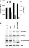
Figure 6. Camptothecin paradoxically increases bioluminescence and luciferase expression.
(A) Effects of camptothecin and 5-azacytidine on bioluminescence of MPC GL-9 cells compared to survival measured by Absorbance in XTT assay at 1 week. (B) corresponding immunoblot from the same experiment showing increased levels of firefly luciferase protein and copepod GFP (copGFP) in camptothecin-treated cultures. Expression of CgA is not increased, indicating that the effect is specific for the luciferase construct. The paradoxically increased bioluminescence of cells treated with camptothecin obscured obvious actual toxicity that was quantifiable by XTT assay.
Figure 7
Figure 7. Clinically utilized TOP1 inhibitors show variable toxicities to MPC cells.
Concurrent tests of camptothecin versus clinically utilized TOP1 inhibitors on MPC cells in monolayer cultures at one week. Equivalency of camptothecin and SN-38 is seen at 10-fold lower concentrations of SN38. Data are from three independent experiments, each with triplicate wells. Bars indicate mean +/− SEM. (**, p<.01; *, p<.05).
Figure 8
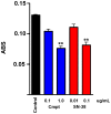
Figure 8. The active metabolite of irinotecan is toxic to primary human pheochromocytoma cells.
XTT assay results showing killing of human PCC cells by camptothecin and SN-38. Cultures were treated with the indicated drug concentrations for one week. Data represent mean +/− SEM of triplicate wells. (**, p<.01).

References

    1. DeLellis RA, Lloyd RV, Heitz PU, Eng C, editors (2004) Tumours of Endocrine Organs. Lyon: IARC Press.
    1. Pacak K, Eisenhofer G, Ahlman H, Bornstein SR, Gimenez-Roqueplo AP, et al. (2007) Pheochromocytoma: recommendations for clinical practice from the First International Symposium. October 2005. Nat Clin Pract Endocrinol Metab 3: 92–102. - PubMed
    1. Strong VE, Kennedy T, Al-Ahmadie H, Tang L, Coleman J, et al. (2008) Prognostic indicators of malignancy in adrenal pheochromocytomas: clinical, histopathologic, and cell cycle/apoptosis gene expression analysis. Surgery 143: 759–768. - PubMed
    1. Tischler AS, Powers JF, Alroy J (2004) Animal models of pheochromocytoma. Histol Histopathol 19: 883–895. - PubMed
    1. Ghayee HK, Bhagwandin VJ, Stastny V, Click A, Ding LH, et al. (2013) Progenitor cell line (hPheo1) derived from a human pheochromocytoma tumor. PLoS One 8: e65624. - PMC - PubMed

Publication types

MeSH terms