

Memory networks in tinnitus: a functional brain image study
- PMID: 24516567
- PMCID: PMC3916334
- DOI: 10.1371/journal.pone.0087839
Memory networks in tinnitus: a functional brain image study
Abstract
Tinnitus is characterized by the perception of sound in the absence of an external auditory stimulus. The network connectivity of auditory and non-auditory brain structures associated with emotion, memory and attention are functionally altered in debilitating tinnitus. Current studies suggest that tinnitus results from neuroplastic changes in the frontal and limbic temporal regions. The objective of this study was to use Single-Photon Emission Computed Tomography (SPECT) to evaluate changes in the cerebral blood flow in tinnitus patients with normal hearing compared with healthy controls.
Methods: Twenty tinnitus patients with normal hearing and 17 healthy controls, matched for sex, age and years of education, were subjected to Single Photon Emission Computed Tomography using the radiotracer ethylenedicysteine diethyl ester, labeled with Technetium 99 m (99 mTc-ECD SPECT). The severity of tinnitus was assessed using the "Tinnitus Handicap Inventory" (THI). The images were processed and analyzed using "Statistical Parametric Mapping" (SPM8).
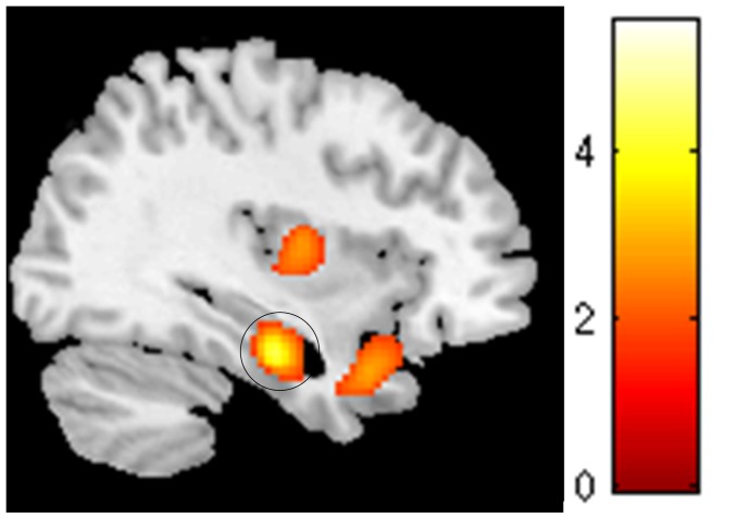
Results: A significant increase in cerebral perfusion in the left parahippocampal gyrus (pFWE <0.05) was observed in patients with tinnitus compared with healthy controls. The average total THI score was 50.8+18.24, classified as moderate tinnitus.
Conclusion: It was possible to identify significant changes in the limbic system of the brain perfusion in tinnitus patients with normal hearing, suggesting that central mechanisms, not specific to the auditory pathway, are involved in the pathophysiology of symptoms, even in the absence of clinically diagnosed peripheral changes.
Conflict of interest statement
Figures
References
-
- Eggermont JJ, Roberts LE (2004) The neuroscience of tinnitus. TRENDS in Neurosciences 27(11): 676–682. - PubMed
-
- Jastreboff PJ (1990) Phantom auditory perception (tinnitus): mechanisms of generation and perception. Neurosci Res 8: 221–254. - PubMed
-
- Gardner A, Pagani M, Jacobsson H, Lindberg G, Larsson SA, et al. (2002) Differences in resting state regional cerebral blood flow assessed with 99mTc-HMPAO SPECT and brain atlas matching between depressed patients with and without tinnitus. Nuclear Medicine Communications 23: 429–439. - PubMed
-
- Noreña AJ, Farley BJ (2013) Tinnitus-related neural activity: theories of generation, propagation, and centralization. Hear Res 295: 161–171. - PubMed
Publication types
MeSH terms
LinkOut - more resources
Full Text Sources
Other Literature Sources
Medical
