. 2014 Feb 6;9(2):e88162.
doi: 10.1371/journal.pone.0088162. eCollection 2014.

Obscurins: Goliaths and Davids take over non-muscle tissues

Affiliations

Obscurins: Goliaths and Davids take over non-muscle tissues

Maegen A Ackermann et al. PLoS One. .

Erratum in

Abstract

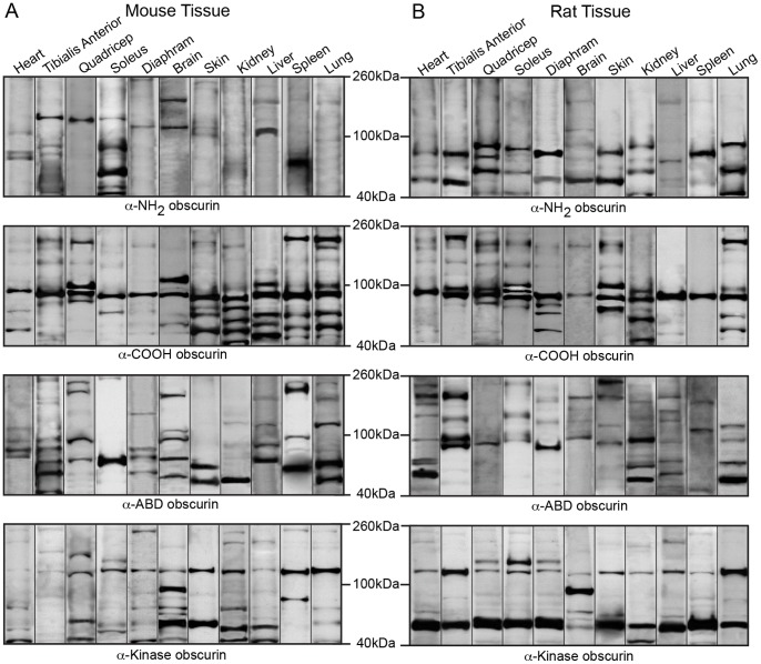
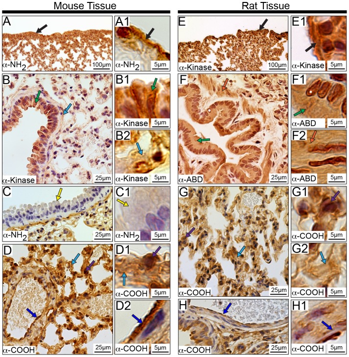
Obscurins comprise a family of proteins originally identified in striated muscles, where they play essential roles in myofibrillogenesis, cytoskeletal organization, and Ca(2+) homeostasis. They are encoded by the single OBSCN gene, and are composed of tandem adhesion domains and signaling motifs. To date, two giant obscurin isoforms have been described in detail that differ only at the extreme COOH-terminus; while obscurin-A (∼720 kDa) contains a non-modular COOH-terminus that harbors binding sites for the adaptor proteins ankyrins, obscurin-B (∼870 kDa) contains two COOH-terminal serine-threonine kinase domains preceded by adhesion motifs. Besides the two known giant obscurins, a thorough search of transcript databases suggests that complex alternative splicing of the obscurin transcript results in the generation of additional giant as well as small isoforms with molecular masses ranging between ∼50-970 kDa. These novel isoforms share common domains with the characterized isoforms, but also contain unique regions. Using a panel of highly specific antibodies directed against epitopes spanning the entire length of giant obscurins, we employed western blotting and immunohistochemistry to perform a systematic and comprehensive characterization of the expression profile of obscurins in muscle and non-muscle tissues. Our studies demonstrate for the first time that obscurins are not restricted to striated muscles, but are abundantly expressed in several tissues and organs including brain, skin, kidney, liver, spleen, and lung. While some obscurin isoforms are ubiquitously expressed, others are preferentially present in specific tissues and organs. Moreover, obscurins are present in select structures and cell types where they assume nuclear, cytosolic, and membrane distributions. Given the ubiquitous expression of some obscurins, along with the preferential expression of others, it becomes apparent that obscurins may play common and unique roles, respectively, in the regulation and maintenance of cell homeostasis in various tissues and organs throughout the body.


Conflict of interest statement

Competing Interests: The authors have declared that no competing interests exist.

Figures

Figure 1
Figure 1. Mammalian obscurin variants.
Domain architecture of up-to-date mammalian obscurin variants as listed in NCBI and Ensembl, illustrating their structural and signaling motifs (please see key for notations). Alternative splicing of the obscurin transcript results in several variants. (A) Obscurin-A-like isoforms, similar to prototypical obscurin-A, containing the non-modular COOH-terminus including the ankyrin-binding domain (ABD). (B) Obscurin-B-like isoforms containing one or both kinase domains, found in the COOH-terminus of obscurin-B. (C) Other splice variants containing sequences specific to neither obscurin-A-like nor obscurin-B-like proteins. The antigenic sequences used for the generation of the four obscurin antibodies are highlighted by the colored boxed regions (α-NH2 in red, α-COOH in blue, α-ABD in green, and α-kinase in yellow; the accession numbers that correspond to the amino acid coordinates of the antigenic sequences are stated in the Materials and Methods section).
Figure 2
Figure 2. Expression of giant obscurins in rodent tissues and organs.
Western blot analysis of 70 µg of protein homogenates prepared from adult mouse (A) and rat (B) tissues were probed with four antibodies to obscurins: α-NH2, α-COOH, α-ABD, and α-Kinase (the epitope for each antibody is noted in Figure 1). The blots have been cut at ∼600 kDa to focus on the giant forms of obscurin, and representative lanes from multiple experiments are shown. In agreement with previously published data, giant obscurin-A and -B are consistently identified in both cardiac and skeletal muscles of mouse and rat origin using any of the four obscurin antibodies. Notably, they are also present in select non-muscle tissues including brain, skin, and lung. Similar protein content per lane was ensured with a GAPDH load control.
Figure 3
Figure 3. Epitopes present in giant obscurins.
The ability of each of the four obscurin antibodies (α-NH2 in red, α-COOH in blue, α-ABD in green, and α-Kinase in yellow) to recognize giant obscurins (>60 kDa) is depicted for each murine tissue and organ.
Figure 4
Figure 4. Expression of intermediate obscurins in rodent tissues and organs.
Western blot analysis of 70 µg of protein homogenates prepared from various adult mouse (A) and rat (B) tissues were probed with antibodies specific to obscurins. As before, probing for GAPDH ensured equal loading. The blots have been cut to include intermediate obscurins, ranging in size between ∼260–600 kDa. Each lane is a representative image from multiple replicates.
Figure 5
Figure 5. Epitopes present in intermediate obscurins.
The ability of each of the four obscurin antibodies (α-NH2 in red, α-COOH in blue, α-ABD in green, and α-Kinase in yellow) to recognize intermediate obscurins (∼260–600 kDa) is noted for each murine tissue and organ.
Figure 6
Figure 6. Expression of small obscurins in rodent tissues and organs.
Western blot analysis of 70 µg of protein homogenates prepared from various adult mouse (A) and rat (B) tissues were probed with antibodies specific to obscurins and a GAPDH loading control. The blots have been cut to show small obscurins with molecular weights of ∼40–260 kDa. A representative blot for each tissue is shown in every lane.
Figure 7
Figure 7. Epitopes present in small obscurins.
The ability of each of the four obscurin antibodies (α-NH2 in red, α-COOH in blue, α-ABD in green, and α-Kinase in yellow) to recognize small obscurins (∼40–260 kDa) is depicted for each murine tissue and organ.
Figure 8
Figure 8. Localization of obscurins in rodent striated muscles.
Adult mouse and rat heart (A-H2) and tibialis anterior (I-N1) muscle sections were analyzed by immunohistochemistry using antibodies specific for obscurins. In accordance with previous studies, obscurins exhibited a striated pattern in both cardiac and skeletal muscles of mouse and rat origin. Cardiac Tissue: Obscurins reside in the mouse and rat epicardium (A-A1 and E-E1, respectively; black arrows). In the myocardial layer, obscurins are found at the sarcolemma (B-B1 and G-G1, mouse and rat tissues, respectively; green arrows) and in sarcomeric striations (C-C1 and F-F1, mouse and rat tissues, respectively; light blue arrows). Interestingly, intercalated disks (pink arrows) and the nuclei of cardiomyocytes (black asterisk) are also labeled in both mouse (D-D1) and rat (H-H2) tissues. In addition, obscurins are found within the cells lining the vasculature throughout the heart (B and B2, and F and F2, mouse and rat tissues, respectively; dark blue arrows). Skeletal Muscle Tissue: Similar to earlier observations, obscurins localize to myofibrillar striations (light blue arrows) of both mouse (I-I1) and rat (L-L1) tissues. Obscurins are also present at the sarcolemma (J-J1 and M-M1, mouse and rat tissues, respectively; green arrows) and the nuclei of mouse, but not rat, skeletal muscle using the α-ABD antibody (K and K2, black asterisk). Moreover, obscurins are found within the cells lining the walls of the vasculature (dark blue arrow) in both mouse (K-K1) and rat (N-N1) tissue. Images are shown at multiple magnifications to highlight the various immunopositive structures. Scale bars are included in each panel for reference; for details please refer to the relevant section in the Materials and Methods.
Figure 9
Figure 9. Distribution of obscurins in the rodent brain.
Obscurins localize to the arachnoid (dark blue arrows) and pia mater (dark green arrows) in both mouse (A-A2) and rat (G-G2) tissue. In addition, obscurins are present in the cell bodies of neurons (black arrow) and the neuropil (orange arrows) of the mouse (B-B1) and rat (H-H1) brain. In the hippocampus, obscurins concentrate in the cytoplasm (yellow arrows) of pyramidal cells of the Cornu Ammonis (CA) both in mouse (C) and rat (I). Additionally, obscurins reside in the cytoplasm (light green arrow) of granule cells of the Dentate Gyrus (DG) of both mouse (D-D1) and rat (J-J1) tissue. Also, in the cerebellum, obscurins are present in the Purkinje cells (light blue arrows) in both mouse (E-E1) and rat (K-K1). Interestingly, fissures within the mouse (F-F1) and rat (L-L1) brain are only labeled with α-NH2 antibody (pink arrows). Images are shown at multiple magnifications to highlight the various immunopositive structures. Scale bars are included in each panel for reference.
Figure 10
Figure 10. Distribution of obscurins in mouse and rat skin.
Obscurins localize to the epidermis (black arrows) of mouse (A-A1) and rat (C-C1) skin. They are also found in the cytoplasm (pink arrows) and nuclei (white asterisks) of epithelial cells composing the root sheath of the hair follicle as well as the cytoplasm (green arrows) and nuclei (white asterisks) of the cells within the sebaceous glands in both mouse (B-B2) and rat (D-D2) tissue. In addition, obscurins are present in the connective tissue (purple arrows) and cells within the connective tissue (yellow arrows) in mouse (B and B3) and rat (D and D3) skin. Similar to the vasculature of striated muscles, obscurins reside within the vasculature of mouse and rat skin (B and B4, and D and D4, respectively; dark blue arrows). Images are shown at multiple magnifications to highlight the various immunopositive structures. Scale bars are included in each panel for reference.
Figure 11
Figure 11. Distribution of obscurins in murine kidney.
In agreement with the other tissues and organs examined, obscurins localize to the outer capsule surrounding the kidney (black arrows) in both mouse (A-A1) and rat (G-G1). Moreover, in the mouse (B and B2) and rat (H and H2) glomerulus, obscurins localize to the cytoplasm (dark green arrows) and nuclei (white asterisks) of endothelial cells. They are also found within the epithelial cells of Bowman’s capsule (B-B1 and H-H1, mouse and rat tissues, respectively; white arrows). Obscurins are also found in the cytoplasm and nuclei (white asterisks) of epithelial cells making up the proximal (pink arrows) and distal (orange arrows) tubules in both mouse (C-C2) and rat (I-I2). Notably, they are present in both the apical (light green arrows) and basolateral (yellow arrow) surfaces of the epithelial cells within distal tubules of mouse (D-E1) and rat (J-K1) tissues. Similarly, they are expressed at the basolateral surface of mouse and rat proximal tubule epithelial cells (E and E2, and K and K2, respectively; light blue arrow), with some accumulation at the apical surface in rat tissues only (J and J2, purple arrows). Obscurins also localize to the vasculature of mouse (F-F1) and rat (L-L1) kidney within both VECs (dark blue arrows) and VSMCs (red arrows). Images are shown at multiple magnifications to highlight the various immunopositive structures. Scale bars are included in each panel for reference.
Figure 12
Figure 12. Localization of obscurins in the rodent liver.
Obscurins localize to the outer surface of the liver, namely Gilsson’s capsule (black arrows) in both mouse (A-A1) and rat (F-F1) tissues. Interestingly, only epitopes at the NH2-terminus of obscurins are found within the connective tissue (yellow arrows) in both mouse (B) and rat (G). Obscurins are also found lining the sinusoids (C-C1, D, and H-H1, mouse and rat tissues, respectively; pink arrows) as well as within the cytoplasm of Kuppfer cells (C, C2, D and E, and H, I, and I2, mouse and rat, respectively; purple arrows) and hepatocytes (C, D, D2, E, and E2 and H, I-I1, J, and J2, mouse and rat, respectively; light blue arrows). Moreover, they are localized to the cell-cell contacts of hepatocytes (green arrows) within both mouse (D-D1) and rat (H and H2) tissue and hepatocyte nuclei (E and E2, and I-I1, mouse and rat respectively; white asterisks). Similar to other tissues and organs, obscurins reside within the VECs (dark blue arrows) of the liver vasculature in both mouse (E-E1) and rat (J-J1). Images are shown at multiple magnifications to highlight the various immunopositive structures. Scale bars are included in each panel for reference.
Figure 13
Figure 13. Distribution of obscurins in the rodent spleen.
Obscurins are present in the outer capsule (black arrows) of the mouse (A-A1) and rat (F-F1) spleen. In addition, they are expressed in the lymphocytes of B-cell follicles of the white pulp (yellow arrows) in both mouse (B) and rat (G). However, obscurins are absent from the T-cell area (purple arrows) surrounding the central arteriole of either mouse (B) or rat (G) spleen. Only those obscurins carrying NH2-terminal epitopes are found within the perivascular region (pink arrows) of both mouse (C) and rat (I) spleen. Unique to rat spleen (H), the cells within the marginal zone surrounding the white pulp are immunopositive for obscurins carrying the α-NH2 and α-kinase epitopes (light blue arrows). Moreover, obscurins are detected in both the cytoplasm (green arrows) and nuclei (white asterisks) of cells residing in the red pulp of both mouse (D-D1) and rat (J-J1) spleen. Obscurins are also present within the trabeculae (orange arrows) of both mouse (D and D2) and rat (J and J2). Furthermore, we observe staining of VSMCs (red arrows) and VECs (dark blue arrows) in both mouse (E-E1) and rat (K-K1) spleen. Images are shown at multiple magnifications to highlight the various immunopositive structures. Scale bars are included in each panel for reference.
Figure 14
Figure 14. Distribution of obscurins in rodent lung.
Obscurins localize to the mesothelial cells making up the pleura, which surrounds the lung (A-A1 and E-E1, mouse and rat, respectively; black arrows). Within the bronchioles, particular obscurins are found in the cytoplasm of Clara cells (green arrows) of both mouse (B-B1) and rat (F-F1) lung. Interestingly, obscurins carrying the NH2-terminal epitopes are absent from the cytoplasm of Clara cells in mouse tissue (C-C1, yellow arrows). Obscurins are also found in the fibrous component of the connective tissue (B, B2, D-D1, G, and G2, light blue arrows) as well as in cells within the connective tissue (purple arrow) in both mouse (D-D1) and rat (G-G1) lung. Similar to observations in other tissues, obscurins localize to VECs (dark blue arrows) in the mouse (D and D2) and rat (H-H1) lung. Smooth muscle within surrounding the bronchioles contains obscurins only in rat tissue (F and F2, orange arrows). Images are shown at multiple magnifications to highlight the various immunopositive structures. Scale bars are included in each panel for reference.

References

    1. Young P, Ehler E, Gautel M (2001) Obscurin, a giant sarcomeric Rho guanine nucleotide exchange factor protein involved in sarcomere assembly. J Cell Biol 154: 123–136. - PMC - PubMed
    1. Russell M (2002) Identification, tissue expression and chromosomal localization of human Obscurin-MLCK, a member of the titin and Dbl families of myosin light chain kinases. Gene 282: 237–246. - PubMed
    1. Kontrogianni-Konstantopoulos A, Bloch RJ (2005) Obscurin: a multitasking muscle giant. J Muscle Res Cell Motil 26: 419–426. - PubMed
    1. Fukuzawa A, Idowu S, Gautel M (2005) Complete human gene structure of obscurin: implications for isoform generation by differential splicing. J Muscle Res Cell Motil 26: 427–434. - PubMed
    1. Perry NA, Ackermann MA, Shriver M, Hu LY, Kontrogianni-Konstantopoulos A (2013) Obscurins: Unassuming giants enter the spotlight. IUBMB Life. - PMC - PubMed

Publication types

LinkOut - more resources