. 2014 Feb 6;9(2):e88193.
doi: 10.1371/journal.pone.0088193. eCollection 2014.

Exosome release and low pH belong to a framework of resistance of human melanoma cells to cisplatin

Affiliations

Exosome release and low pH belong to a framework of resistance of human melanoma cells to cisplatin

Cristina Federici et al. PLoS One. .

Abstract

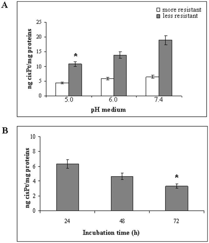
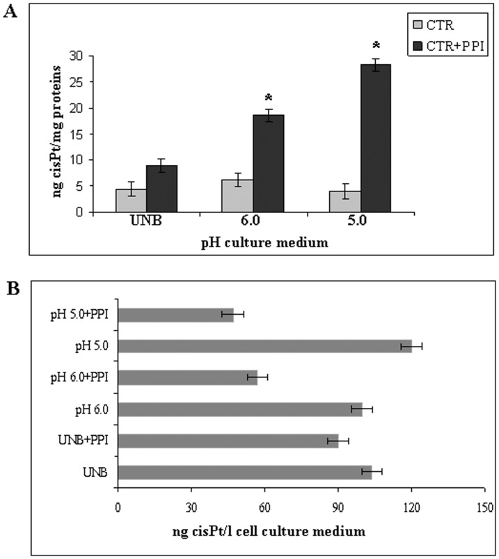
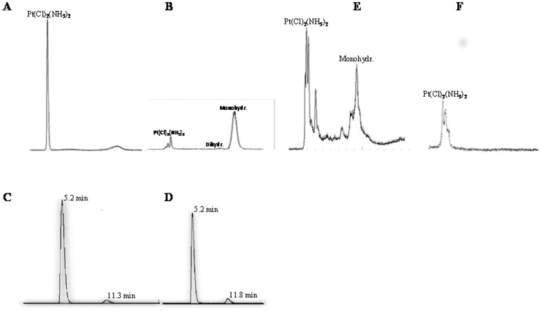
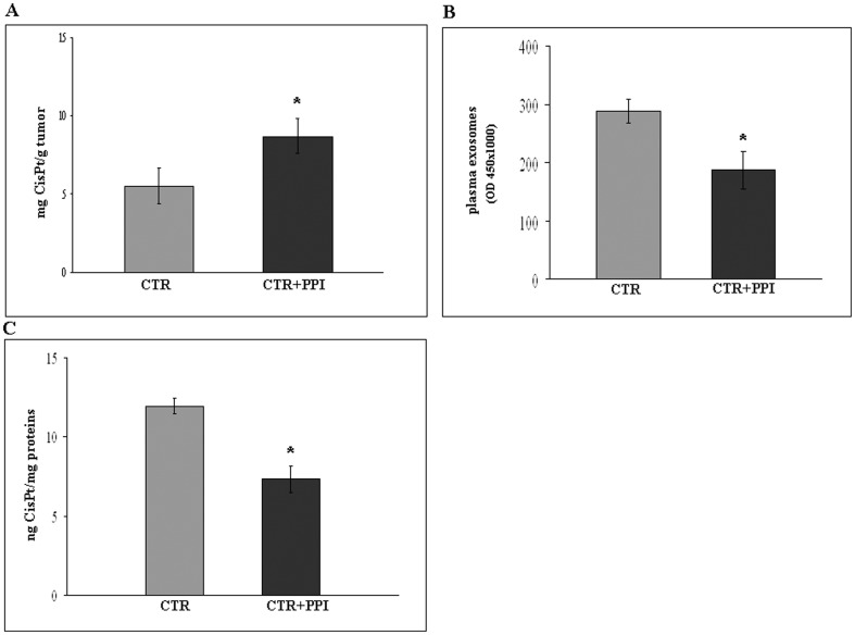
Intrinsic resistance to cytotoxic drugs has been a main issue in cancer therapy for decades. Microenvironmental acidity is a simple while highly efficient mechanism of chemoresistance, exploited through impairment of drug delivery. The latter is achieved by extracellular protonation and/or sequestration into acidic vesicles. This study investigates the importance of extracellular acidosis and nanovesicle (exosome) release in the resistance of human tumour cell to cisplatin (CisPt); in parallel to proton pump inhibitors (PPI) ability of interfering with these tumour cell features. The results showed that CisPt uptake by human tumour cells was markedly impaired by low pH conditions. Moreover, exosomes purified from supernatants of these cell cultures contained various amounts of CisPt, which correlated to the pH conditions of the culture medium. HPLC-Q-ICP-MS analysis revealed that exosome purified from tumour cell culture supernatants contained CisPt in its native form. PPI pre-treatment increased cellular uptake of CisPt, as compared to untreated cells, in an acidic-depend manner. Furthermore, it induced a clear inhibition of exosome release by tumour cells. Human tumours obtained from xenografts pretreated with PPI contained more CisPt as compared to tumours from xenografts treated with CisPt alone. Further analysis showed that in vivo PPI treatment induced a clear reduction in the plasmatic levels of tumour-derived exosomes which also contained lower level of CisPt. Altogether, these findings point to the identification of a double mechanism that human malignant melanoma use in resisting to a dreadful cellular poison such as cisplatin. This framework of resistance includes both low pH-dependent extracellular sequestration and an exosome-mediated elimination. Both mechanisms are markedly impaired by proton pump inhibition, leading to an increased CisPt-dependent cytotoxicity.


Conflict of interest statement

Competing Interests: Stefano Fais has read the journal's policy and the authors have the following conflicts: Patent: A NEW METHOD TO MEASURE AND CHARACTERIZE MICROVESICLES IN THE HUMAN BODY FLUIDS, PCT/EE2009/000001 for the ExoTest described in this work by Hansabiomed Estonia. Inventors: Stefano Fais and Mariantonia Logozzi confirm that their patent (A new method to measure and characterize microvesicles in the human body fluids, PCT/EE2009/000001) doesn't alter the adherence to all the PLOS ONE policies on sharing data and materials, as detailed online in the guide.

Figures

Figure 1
Figure 1. Cytotoxicity assay by Trypan blue exclusion method.
Me30966, Me501, SW480 and MCF7 cell lines were incubated at pH µM of CisPt. Representative of three independent experiments are reported.
Figure 2
Figure 2. Analysis of intracellular CisPt at different pH.
A: Intracellular CisPt level in more drug-resistant (Me30966) and less drug-resistant (MCF7) cells at different pH (5.0, 6.0 and 7.4) of culture medium. Significance (p<0.05) refers to CisPt level at pH 5.0 compared to pH 7.4. B: Intracellular CisPt level in Me30966 cells in function of different incubation times (24, 48, 72 hours) in UNB condition before drug administration. Significance (p<0.05) refers to CisPt uptake after 72 hours compared to 24 hours. Representative of three independent experiments are reported.
Figure 3
Figure 3. Effect of PPI on CisPt cellular uptake.
A: Effect of PPI on CisPt uptake in Me30966 cells in function of different pH (UNB, 6.0 and 5.0) culture medium. CTR: Me30966 cells incubated with CisPt, without PPI pre-treatment; CTR+PPI: Me30966 cells pretreated with PPI and then incubated with CisPt. Significance (p<0.05) refers to CisPt cellular uptake at 5.0 and 6.0 pH in comparing PPI pretreatment to CTR in UNB medium. B: Effect of PPI on drug release at different pH (UNB, 6.0 and 5.0). CisPt ng/l present in cell culture medium obtained from cells pretreated with PPI and then incubated with CisPt. p<0.05. Representative of three independent experiments are reported.
Figure 4
Figure 4. HPLC-Q-ICP-MS chromatograms of a standard solution of CisPt.
Chromatogram of CisPt solution dissolved in NaCl 0.9% (A) and in water after sonication (30 min) at 80°C (90 min) (B). Chromatograms of CisPt dissolved in cell culture medium after dissolution (C) and after 6 hours incubation (D), peak of native form of Cis-Pt at 5.2 min; peak of monohydrated CisPt at 11.3 min. Chromatogram of Me30966 cells lysate solution containing native and monohydrated forms of CisPt (E); chromatogram of exosomes lysate solution containing only the native form of the drug (F). Representative of three independent experiments are reported.
Figure 5
Figure 5. In vivo effect of PPI on CisPt uptake.
A: Effect of PPI (25 mg/kg) on CisPt uptake by human tumour in SCID mice model. CTR: SCID mice treated with CisPt (0,1 mg/mouse), CTR+PPI: SCID mice pretreated with PPI and then with CisPt. p<0.05. B: Effect of PPI in SCID mice plasma on exosomes tumour release analysed by Exo Test. p<0.05. C: Effect of PPI on CisPt content in exosomes circulating in SCID mice. CTR: plasma exosomes of SCID mice treated with CisPt, CTR+PPI: plasma exosomes of SCID mice pretreated with PPI and then with CisPt. p<0.05. Representative of three independent experiments are reported.

Similar articles

Cited by

References

    1. Gray-Schopfer V, Wellbrock C, Marais R (2007) Melanoma biology and new targeted therapy. Nature 22: 851–857. - PubMed
    1. Gogas HJ, Kirkwood JM, Sondak VK (2007) Chemotherapy for metastatic melanoma: time for a change? Cancer 109: 455–464. - PubMed
    1. Chapman PB, Einhorn LH, Meyers ML, Saxman S, Destro AN, et al. (1999) Phase III multicenter randomized trial of the Dartmouth regimen versus dacarbazine in patients with metastatic melanoma. J Clin Oncol 17: 2745–2751. - PubMed
    1. Tsao H, Atkins MB, Sober AJ (2004) Management of cutaneous melanoma. N Engl J Med 351: 998–1012. - PubMed
    1. Ichihashi N, Kitajima Y (2001) Chemotherapy induces or increases expression of multidrug resistance-associated protein in malignant melanoma cells. Br J Dermatol 144: 745–750. - PubMed

MeSH terms