Review
. 2014 Jan;3(1):105-16.
doi: 10.3978/j.issn.2225-319X.2013.12.11.

Illustrated techniques for performing the Cox-Maze IV procedure through a right mini-thoracotomy

Affiliations
Review

Illustrated techniques for performing the Cox-Maze IV procedure through a right mini-thoracotomy

Jason O Robertson et al. Ann Cardiothorac Surg. 2014 Jan.

Abstract

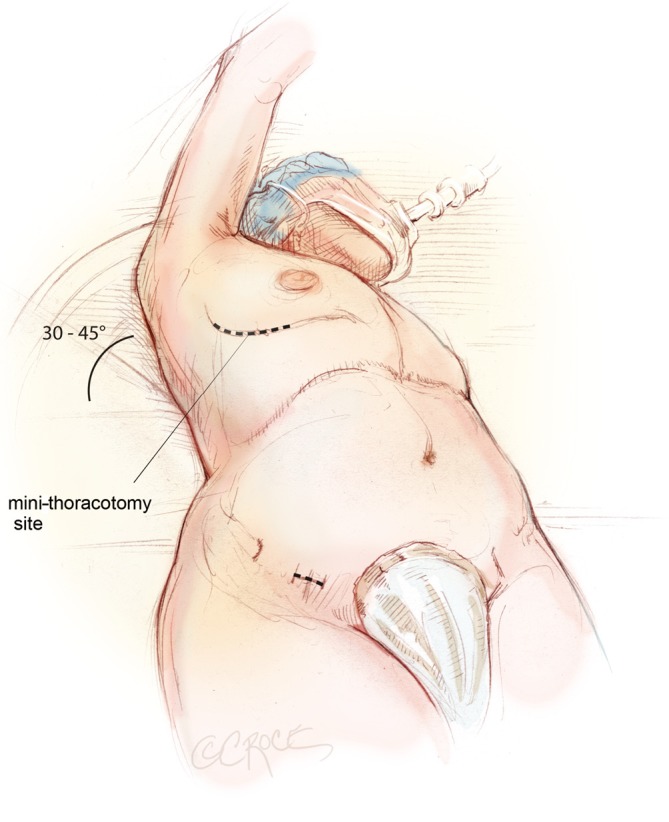
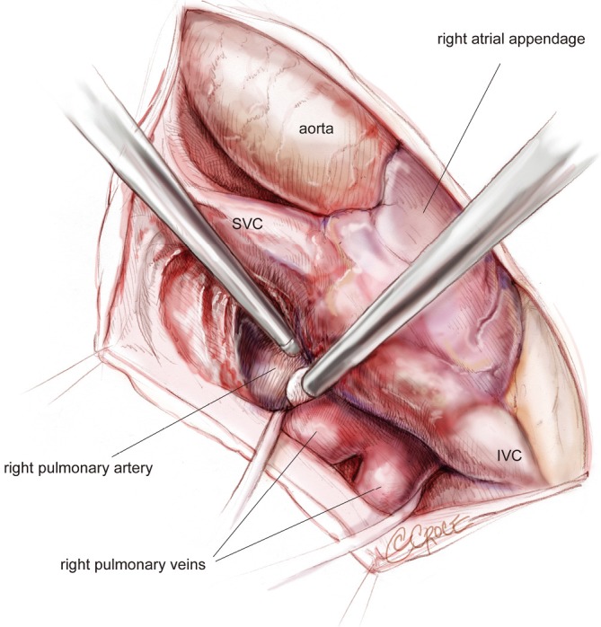
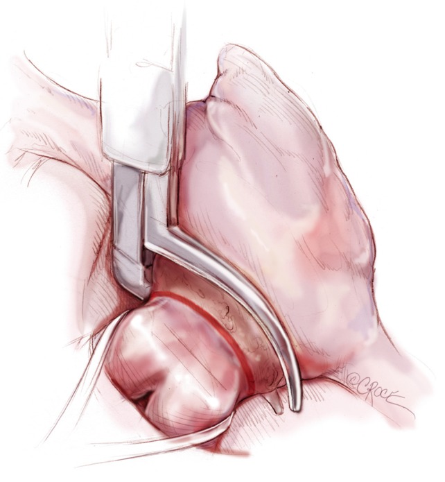
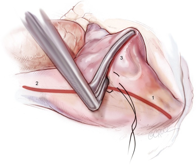
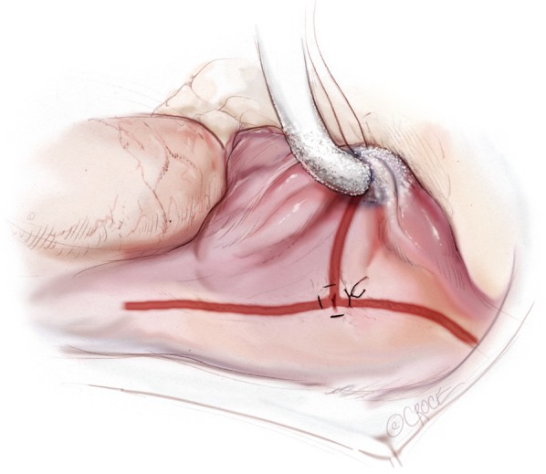
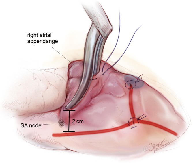
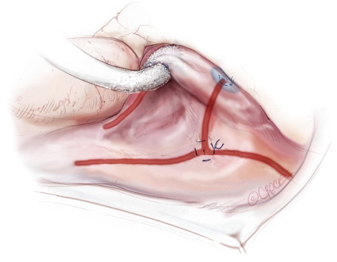
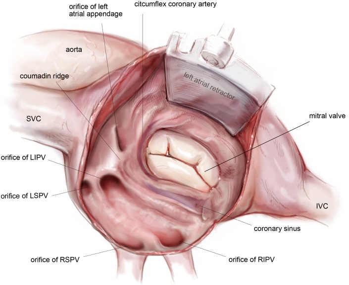
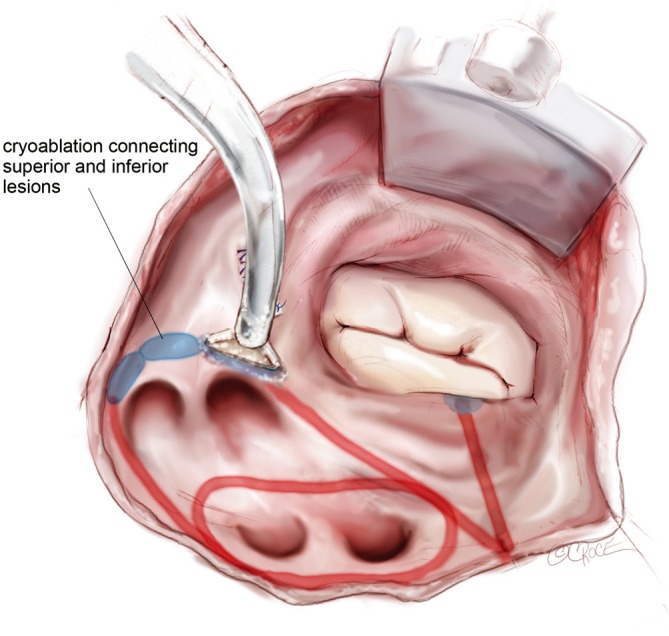
The Cox-Maze IV procedure has replaced the "cut-and-sew" technique of the original Cox-Maze operation with lines of ablation created using bipolar radiofrequency (RF) and cryothermal energy devices. In select patients, this procedure can be performed through a right mini-thoracotomy. This illustrated review is the first to detail the complete steps of the Cox-Maze IV procedure performed through a right mini-thoracotomy with careful attention paid to operative anatomy and advice. Pre- and post-operative management and outcomes are also discussed. This should be a practical guide for the practicing cardiac surgeon.

Keywords: Atrial fibrillation (AF); Cox-Maze IV; right mini-thoracotomy.


Figures

Figure 1
Figure 1
Positioning and marking in preparation for a Cox-Maze IV procedure performed through a right mini-thoracotomy. This depiction of the procedure on a woman demonstrates a submammary incision, as well as a subinguinal incision that is used to dissect the femoral vessels and initiate cardiopulmonary bypass (CPB).
Figure 2
Figure 2
Mobilization of the space between the right superior pulmonary vein and the right pulmonary artery from underneath the SVC, entering into the oblique sinus. SVC, superior vena cava.
Figure 3
Figure 3
Right-sided pulmonary vein isolation using a bipolar RF clamp. RF, radiofrequency.
Figure 4
Figure 4
Ablations of the [1] IVC, [2] SVC and [3] right atrial free wall directed towards the AV groove near the acute margin of the heart. Ablations are performed using a bipolar RF clamp through a purse-string suture placed midway between the SVC and IVC. IVC, inferior vena cava; SVC, superior vena cava; AV, atrioventricular; RF, radiofrequency.
Figure 5
Figure 5
Cryoablation towards the tricuspid annulus at the 1 o’clock position through a second purse-string suture that is placed near the AV groove at the end of the right atrial free wall ablation line. AV, atrioventricular.
Figure 6
Figure 6
Second right atrial free wall ablation performed using a bipolar RF clamp. The clamp is placed through a third purse-string at the base of the RAA. At least two centimeters should be left between this ablation line and the SVC line in order to avoid injury to the sinus node. RF, radiofrequency; SVC, superior vena cava.
Figure 7
Figure 7
Completion of the right atrial lesion set with creation of an endocardial cryoablation to the 11 o’clock position of the tricuspid annulus. This is performed using a linear cryoprobe inserted through the purse-string at the base of the RAA.
Figure 8
Figure 8
Anatomy of the left atrium following an atriotomy. Exposure is obtained using a left atrial lift system. In a left dominant system, the circumflex artery wraps further around the mitral valve, parallel to the coronary sinus. SVC, superior vena cava; LIPV, left inferior pulmonary vein; LSPV, left superior pulmonary vein; RSPV, right superior pulmonary vein; RIPV, right inferior pulmonary vein; IVC, inferior vena cava.
Figure 9
Figure 9
Closure of the left atrial appendage from the endocardial surface of the left atrium. The appendage is oversewn in two layers using a running suture, starting with a pledgeted horizontal mattress stitch at the inferior corner of the orifice.
Figure 10
Figure 10
Ablation to the mitral isthmus using a bipolar RF clamp. The inferior and superior connecting lesions are also depicted after creation with the same device. RF, radiofrequency.
Figure 11
Figure 11
Endocardial cryoablation using a T-shaped cryoprobe to connect the mitral isthmus line to the mitral annulus. The ends of the mitral isthmus and inferior connecting lesions that were created with the bipolar RF clamp are marked with methylene blue in order to provide starting points for this cryoablation, as well as subsequent ones designed to completely isolate the posterior left atrium. RF, radiofrequency.
Figure 12
Figure 12
Epicardial ablation of the coronary sinus in line with the mitral isthmus line using a linear cryoprobe. This is performed with the T-shaped cryoprobe still in place in order to compress the tissue and ensure a full thickness ablation.
Figure 13
Figure 13
A sequence of endocardial cryoablations behind the left pulmonary veins is created with a T-shaped cryoprobe in order to connect the left inferior and superior connecting lesions and ensure complete isolation of the posterior left atrium.

References

    1. Cox JL, Ad N, Palazzo T. Impact of the maze procedure on the stroke rate in patients with atrial fibrillation. J Thorac Cardiovasc Surg 1999;118:833-40 - PubMed
    1. Feinberg MS, Waggoner AD, Kater KM, et al. Restoration of atrial function after the maze procedure for patients with atrial fibrillation. Assessment by Doppler echocardiography. Circulation 1994;90:II285-92 - PubMed
    1. Cox JL, Schuessler RB, Lappas DG, et al. An 8 1/2-year clinical experience with surgery for atrial fibrillation. Ann Surg 1996;224:267-73; discussion 273-5 - PMC - PubMed
    1. Prasad SM, Maniar HS, Camillo CJ, et al. The Cox maze III procedure for atrial fibrillation: long-term efficacy in patients undergoing lone versus concomitant procedures. J Thorac Cardiovasc Surg 2003;126:1822-8 - PubMed
    1. Gammie JS, Haddad M, Milford-Beland S, et al. Atrial fibrillation correction surgery: lessons from the Society of Thoracic Surgeons National Cardiac Database. Ann Thorac Surg 2008;85:909-14 - PubMed