. 2014 Feb;10(2):831-42.
doi: 10.1016/j.actbio.2013.10.020.

Thermally responsive nanoparticle-encapsulated curcumin and its combination with mild hyperthermia for enhanced cancer cell destruction

Thermally responsive nanoparticle-encapsulated curcumin and its combination with mild hyperthermia for enhanced cancer cell destruction

Wei Rao et al. Acta Biomater. 2014 Feb.

Abstract

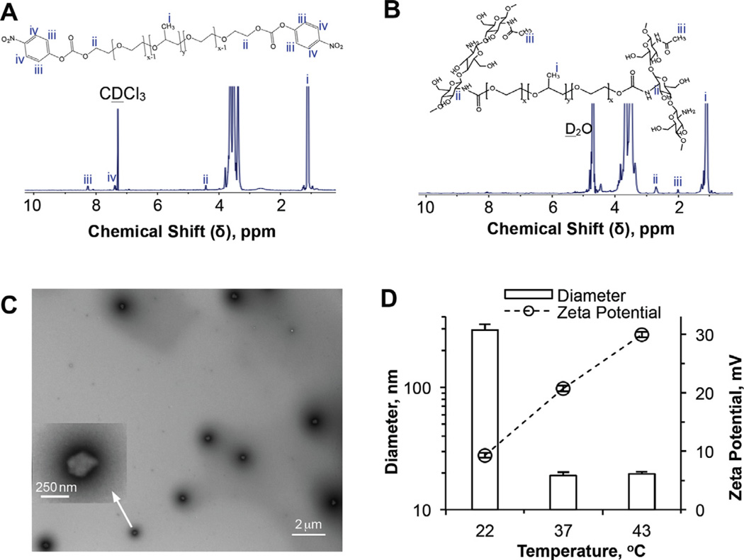
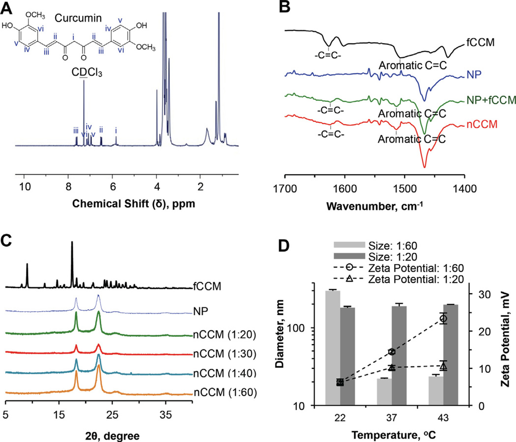
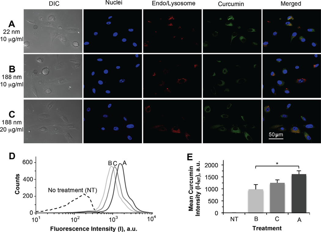
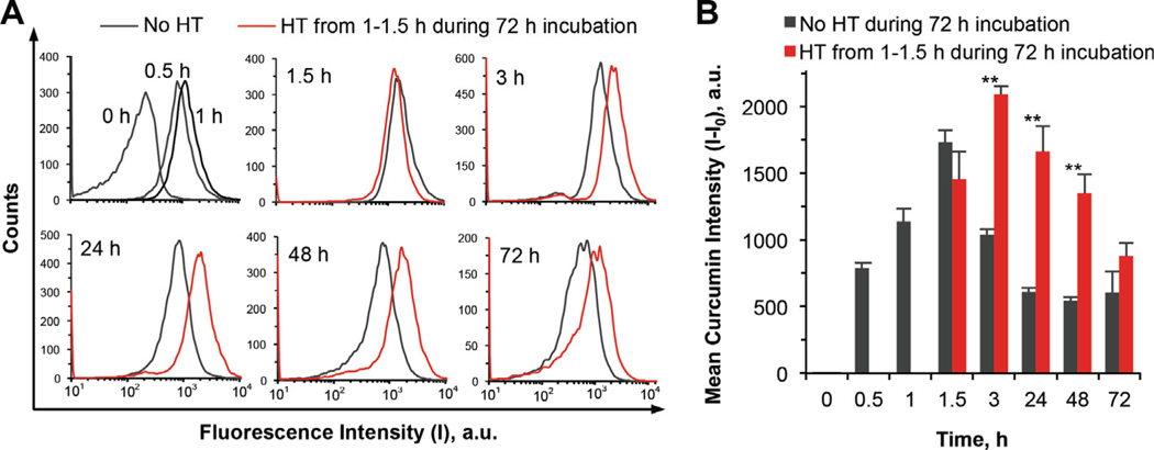
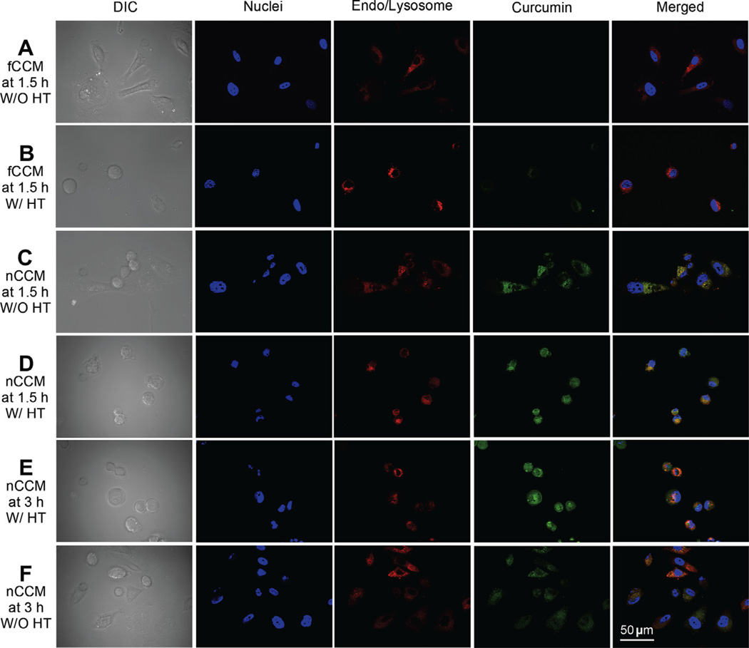
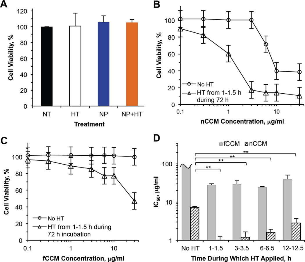
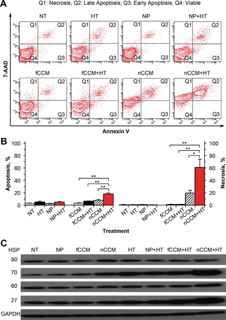
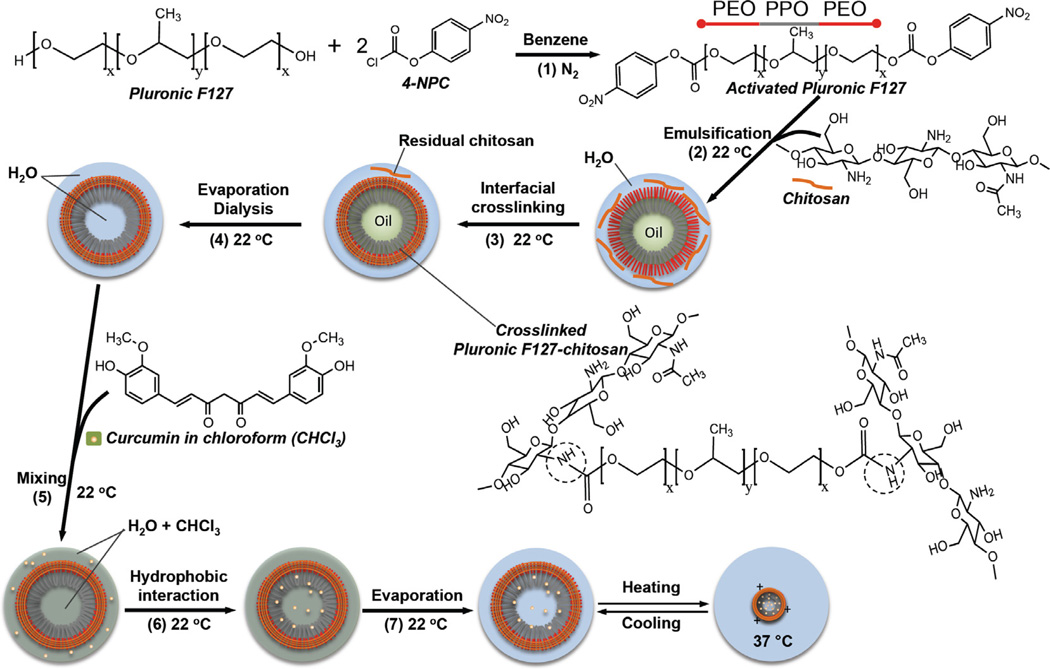
In this study, thermally responsive polymeric nanoparticle-encapsulated curcumin (nCCM) was prepared and characterized. The nCCM is ≈ 22 and 300 nm in diameter at 37 and 22 °C, respectively. The smaller size of the nCCM at 37 °C was found to significantly facilitate its uptake in vitro by human prostate adenocarcinoma PC-3 cancer cells. However, the intracellular nCCM decreases rapidly (rather than plateaus) after reaching its peak at ≈ 1.5 h during a 3-day incubation of the PC-3 cells with nCCM. Moreover, a mild hyperthermia (with negligible cytotoxicity alone) at 43 °C applied between 1 and 1.5 h during the 3-day incubation not only increases the peak uptake but also alters intracellular distribution of nCCM (facilitating its delivery into cell nuclei), which helps to retain a significantly much higher level of intracellular curcumin. These effects of mild hyperthermia could be due in part to the thermal responsiveness of the nCCM: they are more positively charged at 43 °C and can be more easily attracted to the negatively charged nuclear membrane to enter nuclei as a result of electrostatic interaction. Ultimately, a combination of the thermally responsive nCCM and mild hyperthermia significantly enhances the anticancer capability of nCCM, resulting in a more than 7-fold decrease in its inhibitory concentration to reduce cell viability to 50% (IC50). Further mechanistic studies suggest injury pathways associated with heat shock proteins 27 and 70 should contribute to the enhanced cancer cell destruction by inducing cell apoptosis and necrosis. Overall, this study demonstrates the potential of combining mild hyperthermia and thermally responsive nanodrugs such as nCCM for augmented cancer therapy.


Conflict of interest statement

Disclosure

The authors declare no conflict of interests.

Figures

Fig. 1
Fig. 1
Characterization of activated Pluronic F127 and Pluronic F127–chitosan nanoparticles: 1H NMR spectra of (A) 4-NPC activated Pluronic F127 in CDCl3 and (B) Pluronic F127–chitosan nanoparticles in D2O, showing characteristic peaks of 4-NPC and chitosan, respectively; (C) TEM image of Pluronic F127–chitosan nanoparticles showing their core–shell morphology (the inset shows a zoom-in view of one of the nanoparticles); and (D) DLS data showing thermal responsiveness of the nanoparticles in size (diameter) and surface charge (represented by zeta potential).
Fig. 2
Fig. 2
Characterization of nCCM: (A) 1H NMR spectrum showing successful encapsulation of curcumin in Pluronic F127–chitosan nanoparticles to obtain nCCM; (B) FTIR spectra of fCCM, NP, a simple mixture of NP and fCCM (NP + fCCM) and nCCM, showing characteristic peaks due to the aliphatic and aromatic double bonds in curcumin; (C) XRD data showing no clear curcumin peak for nCCM obtained under different feeding ratios of curcumin to nanoparticles, indicating the amorphous state of nCCM in nanoparticles; and (D) DLS data of diameter and surface zeta potential of nCCM obtained with feeding ratios of 1:60 and 1:20.
Fig. 3
Fig. 3
Effect of size (~22 vs. 188 nm) of the nCCM on its uptake by PC-3 cancer cells: (A–C) typical DIC and Apotome SIM fluorescence micrographs; (D) typical flow cytometry peaks of fluorescence intensity (I) in cells treated under the conditions for (A–C), together with that in NT control cells; and (E) a summary of the mean curcumin intensity (IINT) in cells from flow cytometry of three independent runs, showing uptake of nCCM of either ~22 nm at 10 µg ml−1 or ~188 nm at 10 and 20 µg ml−1 in PC-3 cancer cells after 1 h incubation at 37 °C. The cells can take up the smaller (~22 nm) nCCM significantly faster. In (A–C), the blue and red stains are Hoechst and LysoTracker Red staining of cell nuclei and endo/lysosomes, respectively. The latter stains are spot-like under enlarged view. In (E), the mean curcumin intensity was calculated as the difference in fluorescence intensity in the cells with various treatment (I) and that with no treatment (Int) *p< 0.05.
Fig. 4
Fig. 4
Effect of HT on cellular uptake of ~22 nm nCCM: (A) typical flow cytometry peaks of fluorescence intensity in PC-3 cancer cells and (B) mean curcumin intensity (II0) in the cells at various times during incubation of the cells with the nCCM at 37 °C for 72 h either with or without a mild hyperthermia at 43 °C applied from 1 to 1.5 h: the HT treatment not only increases the peak uptake but also helps retain a much higher level of intracellular curcumin from 3 to 72 h. The mean curcumin intensity was calculated as the difference in fluorescence intensity in the cells at various time (I) and that at 0 h (I0). **p < 0.01.
Fig. 5
Fig. 5
Effect of HT on cellular uptake and intracellular distribution of free and ~22 nm nCCM: typical DIC and Apotome SIM fluorescence micrographs of PC-3 cancer cells taken at 1.5 h (A–D) and 3 h (E and F) after incubating the cells with 10 µg ml−1 free curcumin (fCCM, A and B) or ~22 nm nCCM (C–F), either with or without HT applied from 1 to 1.5 h during the incubation. The blue and red stains are Hoechst and LysoTracker Red staining of cell nuclei and endo/lysosomes, respectively, and the latter stains are spot-like under enlarged view.
Fig. 6
Fig. 6
Toxicity to PC-3 cancer cells treated with HT, together with NP, fCCM, nCCM (~22 nm) and their combination with HT: (A) viability at day 3 of PC-3 cancer cells with NT, after treated with HT from 1 to 1.5 h alone, incubated with 1.845 mg ml−1 (corresponding to 30 µg ml−1 nCCM) NP for 3 days alone, and treated with the combination of the 3-day NP incubation and HT from 1 to 1.5 h during the incubation (NP + HT); viability of PC-3 cancer cells after 3-day incubation with ~22 nm nCCM (B) or fCCM (C) of various concentrations either without or with HT applied from 1 to 1.5 h during the incubation; and (D) IC50 (inhibitory concentration to reduce cell viability to 50%) determined using the data shown in (B) and (C) together with that shown in Fig. S5 for cells incubated for 3 days with fCCM and nCCM in combination with HT applied at 3–3.5 h, 6–6.5 h and 12–12.5 h: applying HT at 1–1.5 h during incubation with nCCM gives the best cancer cell destruction for the combined treatment on average, although the difference is statistically insignificant. All treatments were done in culture medium containing 10% FBS. For unit conversion, 1 µg ml−1 is equivalent to 2.71 µM curcumin. All viability data were calculated with respect to control cells with no treatment. The IC50 data for fCCM with HT at 3–3.5, 6–6.5 and 12–12.5 h were obtained with slight linear extrapolation. **p < 0.01.
Fig. 7
Fig. 7
Mechanisms of injury to PC-3 cancer cells after incubated in medium for 2 days with NT, HT alone from 1 to 1.5 h during the incubation, NP alone, NP + HT, fCCM, fCCM + HT, nCCM (~22 nm) or nCCM + HT: (A) typical two-channel (for annexin V and 7-AAD) flow cytometry data showing the distribution of cells with necrosis in quadrant 1 (Q1), late apoptosis in Q2, early apoptosis in Q3 and viable cells in Q4; (B) a summary of the flow cytometry data from three independent runs; and (C) expression of four HSP showing possible injury mechanism involving HSP 70 and 27 for the combined treatment of nCCM and HT. *p < 0.05; **p < 0.01.
Scheme 1
Scheme 1
A schematic illustration of the chemistry and procedure for activating Pluronic F127, synthesizing Pluronic F127–chitosan NP and encapsulating free curcumin (fCCM) in the nanoparticle to obtain nanoparticle-encapsulated curcumin (nCCM): Pluronic F127 was activated (1) at both terminals using 4-NPC; nanoparticles were synthesized by oil-in-water emulsification (2), interfacial crosslinking with chitosan (3), and rotary evaporation and rigorous dialysis (4) to obtain aqueous solution of pure nanoparticles. Curcumin was encapsulated in the nanoparticle by mixing (under constant stirring) (5) solutions of free curcumin (fCCM) in chloroform (CHCl3) and nanoparticles in water, utilizing (6) the high affinity between curcumin and the hydrophobic PPO core of the nanoparticles, and removing (7) chloroform by rotary evaporation. Also shown is an illustration of the thermal responsiveness of the resultant nCCM. The dashed circle in the formula of the crosslinked Pluronic F127–chitosan indicates the bonding between chitosan and Pluronic F127.

References

    1. Rodwell C. Curcumin curries favour? Nat Rev Cancer. 2012;12:376.
    1. Luer S, Troller R, Aebi C. Antibacterial and antiinflammatory kinetics of curcumin as a potential antimucositis agent in cancer patients. Nutr Cancer. 2012;64:975–981. - PubMed
    1. Agrawal DK, Mishra PK. Curcumin and its analogues: potential anticancer agents. Med Res Rev. 2010;30:818–860. - PubMed
    1. Tang H, Murphy CJ, Zhang B, Shen Y, Van Kirk EA, Murdoch WJ, et al. Curcmin polymers as anticancer conjugates. Biomaterials. 2010;31:7139–7149. - PubMed
    1. Sharma RA, Euden SA, Platton SL, Cooke DN, Shafayat A, Hewitt HR, et al. Phase clinical trial of oral curcumin: biomarkers of systemic activity and compliance. Clin Cancer Res. 2004;10:6847–6854. - PubMed

Publication types

LinkOut - more resources