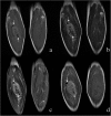
Magnetic resonance imaging features of alveolar soft part sarcoma: report of 14 cases
- PMID: 24517100
- PMCID: PMC3931484
- DOI: 10.1186/1477-7819-12-36
Magnetic resonance imaging features of alveolar soft part sarcoma: report of 14 cases
Abstract
Background: The purpose of this study was to assess the magnetic resonance imaging findings of alveolar soft part sarcoma.
Methods: Magnetic resonance images of pathologically proven alveolar soft part sarcoma in 14 patients were retrospectively reviewed, including lesion location, size and shape, border definition, signals on T1-weighted and T2-weighted images, presence or absence of peritumoral and intratumoral flow voids, and enhancement pattern.
Results: Patients included five women and nine men, ranging in age from 27 to 54 years, with a mean age of 36 years. A slow-growing mass without pain was the chief complaint. Eight patients had pulmonary metastases at presentation. Ten lesions arose from the extremities, two were located in the gluteal regions, one affected the presacral space and one occurred in the back. The mean maximal size of the lesions was 9.8 cm, ranging from 6.2 to 16 cm. All lesions appeared as a round (n = 2), ovoid (n = 8) or irregular (n = 4) shape with ill-defined margins. The lesions mainly demonstrated isointense or mildly hyperintense compared to muscle on T1-weighted images, and heterogeneous high signal intensity on T2-weighted images. Peritumoral edemas were observed in six patients. Ten lesions showed intense inhomogeneous enhancement after contrast. Intra- and peritumoral tubular flow voids representing tortuous dilated vessels with rapid blood flow were present in all cases.
Conclusions: Alveolar soft part sarcoma has some distinctive magnetic resonance imaging features including a slow-growing, large mass in the soft tissue of the extremities in young adults, with numerous signal voids on T1-weighted and T2-weighted images, and strong enhancement after contrast.
Figures


Similar articles
-
Alveolar soft-part sarcoma: can MRI help discriminating from other soft-tissue tumors? A study of the French sarcoma group.Eur Radiol. 2019 Jun;29(6):3170-3182. doi: 10.1007/s00330-018-5903-3. Epub 2018 Dec 17. Eur Radiol. 2019. PMID: 30560359
-
Clinical presentation and CT/MRI findings of alveolar soft part sarcoma: a retrospective single-center analysis of 14 cases.Acta Radiol. 2016 Apr;57(4):475-80. doi: 10.1177/0284185115597720. Epub 2015 Jul 31. Acta Radiol. 2016. PMID: 26231949
-
Magnetic Resonance Features and Characteristic Vascular Pattern of Alveolar Soft-Part Sarcoma.Oncol Res Treat. 2017;40(10):580-585. doi: 10.1159/000477443. Epub 2017 Sep 20. Oncol Res Treat. 2017. PMID: 28950275
-
Magnetic resonance imaging of soft tissue sarcoma: features related to prognosis.Eur J Orthop Surg Traumatol. 2021 Dec;31(8):1567-1575. doi: 10.1007/s00590-021-03003-2. Epub 2021 May 29. Eur J Orthop Surg Traumatol. 2021. PMID: 34052920 Review.
-
Alveolar soft part sarcoma of the uterine cervix in a woman presenting with postmenopausal bleeding: a case report and literature review.Eur J Gynaecol Oncol. 2011;32(3):359-61. Eur J Gynaecol Oncol. 2011. PMID: 21797137 Review.
Cited by
-
Alveolar soft-part sarcoma: can MRI help discriminating from other soft-tissue tumors? A study of the French sarcoma group.Eur Radiol. 2019 Jun;29(6):3170-3182. doi: 10.1007/s00330-018-5903-3. Epub 2018 Dec 17. Eur Radiol. 2019. PMID: 30560359
-
Imaging and Pathological Features of Alveolar Soft Part Sarcoma: Analysis of 16 Patients.Indian J Radiol Imaging. 2021 Sep 7;31(3):573-581. doi: 10.1055/s-0041-1735501. eCollection 2021 Jul. Indian J Radiol Imaging. 2021. PMID: 34790300 Free PMC article.
-
iCREATE: imaging features of primary and metastatic alveolar soft part sarcoma from the EORTC CREATE study.Cancer Imaging. 2020 Oct 30;20(1):79. doi: 10.1186/s40644-020-00352-9. Cancer Imaging. 2020. PMID: 33121537 Free PMC article.
-
The dynamic contrast enhanced-magnetic resonance imaging and diffusion-weighted imaging features of alveolar soft part sarcoma.Quant Imaging Med Surg. 2023 Oct 1;13(10):7269-7280. doi: 10.21037/qims-23-743. Epub 2023 Sep 11. Quant Imaging Med Surg. 2023. PMID: 37869277 Free PMC article.
-
Ultrasound characteristics of alveolar soft part sarcoma in pediatric patients: a retrospective analysis.BMC Cancer. 2024 Dec 2;24(1):1484. doi: 10.1186/s12885-024-13262-x. BMC Cancer. 2024. PMID: 39623317 Free PMC article.
References
-
- Iwamoto Y, Morimoto N, Chuman H, Shinohara N, Sugioka Y. The role of MR imaging in the diagnosis of alveolar soft part sarcoma: a report of 10 cases. Skeletal Radiol. 1995;24:267–270. - PubMed
MeSH terms
LinkOut - more resources
Full Text Sources
Other Literature Sources
Medical

