

Human fibrocytes express multiple antigens associated with autoimmune endocrine diseases
- PMID: 24517144
- PMCID: PMC4010713
- DOI: 10.1210/jc.2013-3072
Human fibrocytes express multiple antigens associated with autoimmune endocrine diseases
Abstract
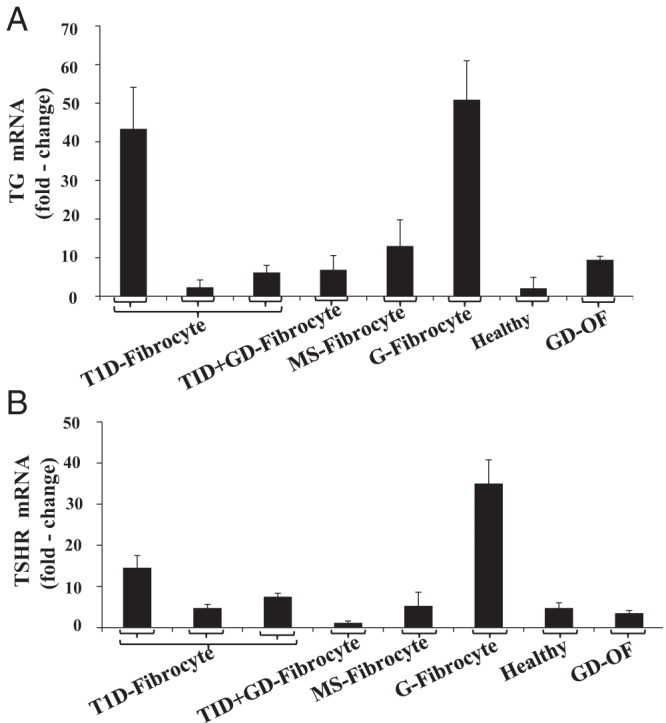
Context: Factors common to multiple autoimmune diseases have been sought vigorously. Graves' disease (GD) and type 1 diabetes mellitus (T1DM) involve end-organ remodeling. Fibrocytes participate in inflammatory diseases and were recently shown to express thyroid-specific proteins such as the thyrotropin receptor and thyroglobulin.
Objective: The objective of the study was to determine whether a broader repertoire of autoantigen expression, such as proteins associated with T1DM, can be ascribed to fibrocytes.
Design, setting, and participants: Fibrocytes and fibroblasts were collected and analyzed from healthy individuals and those with autoimmune diseases in an academic clinical practice.
Main outcome measures: Real-time PCR, Western blot analysis, gene promoter analysis, cell transfections, and flow cytometric cell sorting were performed.
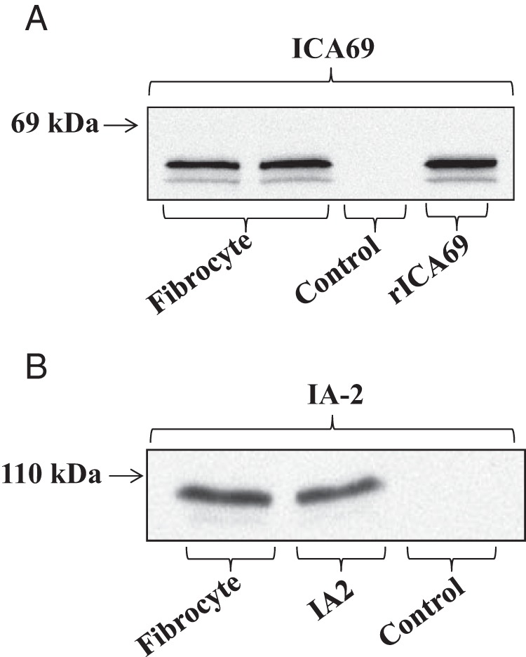
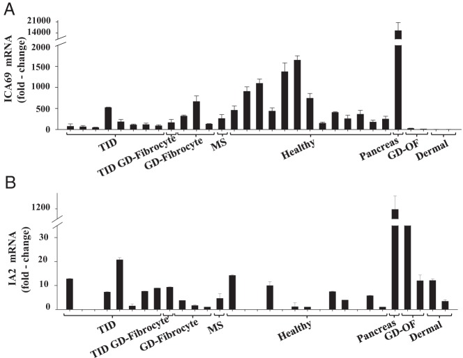
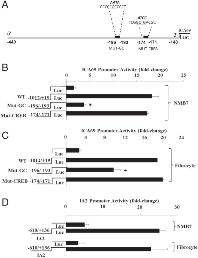
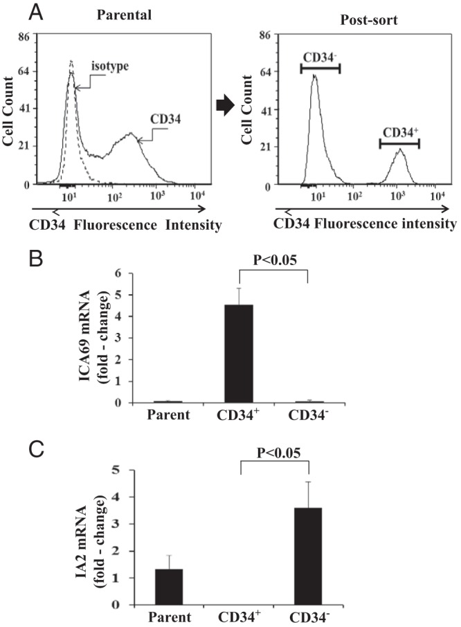
Results: Islet cell antigen ICA512 (IA-2) and islet cell autoantigen of 69 kDa (ICA69), two islet-specific proteins implicated in T1DM, are expressed by fibrocytes from healthy donors and those with T1DM, GD, and multiple sclerosis. Both transcripts are detected by PCR, the proteins are resolved on Western blots, and both gene promoters are active in fibrocytes. Levels of ICA69 are substantially higher than those of IA-2 in fibrocytes. ICA69 localizes to CD34(+) GD orbital fibroblasts putatively derived from fibrocytes, whereas higher levels of IA-2 are found in CD34(-) fibroblasts.
Conclusions: In addition to autoantigens implicated in thyroid autoimmunity, fibrocytes and derivative fibroblasts express multiple autoantigens associated with T1DM. This expression results from active gene promoters and abundant steady-state mRNA encoding ICA69 and IA-2. These latest findings demonstrate that fibrocytes express antigens relevant to multiple forms of endocrine autoimmunity. They suggest the potential for these cells playing a direct role in immune reactivity directed at the thyroid and pancreatic islets.
Figures
Similar articles
-
Potential Roles of CD34+ Fibrocytes Masquerading as Orbital Fibroblasts in Thyroid-Associated Ophthalmopathy.J Clin Endocrinol Metab. 2019 Feb 1;104(2):581-594. doi: 10.1210/jc.2018-01493. J Clin Endocrinol Metab. 2019. PMID: 30445529 Free PMC article. Review.
-
Expression of thyrotropin receptor, thyroglobulin, sodium-iodide symporter, and thyroperoxidase by fibrocytes depends on AIRE.J Clin Endocrinol Metab. 2014 Jul;99(7):E1236-44. doi: 10.1210/jc.2013-4271. Epub 2014 Apr 7. J Clin Endocrinol Metab. 2014. PMID: 24708100 Free PMC article.
-
Fibroblasts expressing the thyrotropin receptor overarch thyroid and orbit in Graves' disease.J Clin Endocrinol Metab. 2011 Dec;96(12):3827-37. doi: 10.1210/jc.2011-1249. Epub 2011 Sep 28. J Clin Endocrinol Metab. 2011. PMID: 21956421 Free PMC article.
-
Slit2 Modulates the Inflammatory Phenotype of Orbit-Infiltrating Fibrocytes in Graves' Disease.J Immunol. 2018 Jun 15;200(12):3942-3949. doi: 10.4049/jimmunol.1800259. Epub 2018 May 11. J Immunol. 2018. PMID: 29752312 Free PMC article.
-
Autoimmune diabetes: the role of T cells, MHC molecules and autoantigens.Autoimmunity. 1998;27(3):159-77. doi: 10.3109/08916939809003864. Autoimmunity. 1998. PMID: 9609134 Review.
Cited by
-
Potential Roles of CD34+ Fibrocytes Masquerading as Orbital Fibroblasts in Thyroid-Associated Ophthalmopathy.J Clin Endocrinol Metab. 2019 Feb 1;104(2):581-594. doi: 10.1210/jc.2018-01493. J Clin Endocrinol Metab. 2019. PMID: 30445529 Free PMC article. Review.
-
Teprotumumab as a Novel Therapy for Thyroid-Associated Ophthalmopathy.Front Endocrinol (Lausanne). 2020 Dec 17;11:610337. doi: 10.3389/fendo.2020.610337. eCollection 2020. Front Endocrinol (Lausanne). 2020. PMID: 33391187 Free PMC article. Review.
-
Pentraxin-3 Is a TSH-Inducible Protein in Human Fibrocytes and Orbital Fibroblasts.Endocrinology. 2015 Nov;156(11):4336-44. doi: 10.1210/en.2015-1399. Epub 2015 Aug 19. Endocrinology. 2015. PMID: 26287404 Free PMC article.
-
IGF1 receptor and thyroid-associated ophthalmopathy.J Mol Endocrinol. 2018 Jul;61(1):T29-T43. doi: 10.1530/JME-17-0276. Epub 2017 Dec 22. J Mol Endocrinol. 2018. PMID: 29273685 Free PMC article. Review.
-
TSH-receptor-expressing fibrocytes and thyroid-associated ophthalmopathy.Nat Rev Endocrinol. 2015 Mar;11(3):171-81. doi: 10.1038/nrendo.2014.226. Epub 2015 Jan 6. Nat Rev Endocrinol. 2015. PMID: 25560705 Free PMC article. Review.
References
-
- Sellner J, Kalluri SR, Cepok S, Hemmer B, Berthele A. Thyroid antibodies in aquaporin 4 antibody positive central nervous system autoimmunity and multiple sclerosis. Clin Endocrinol (Oxf). 2011;75:271–272 - PubMed
-
- Pietropaolo M, Peakman M, Pietropaolo SL, et al. Combined analysis of GAD65 and ICA512(IA-2) autoantibodies in organ and non-organ-specific autoimmune diseases confers high specificity for insulin-dependent diabetes mellitus. J Autoimmun. 1998;11:1–10 - PubMed
-
- Karges W, Hammond-McKibben D, Gaedigk R, Shibuya N, Cheung R, Dosch HM. Loss of self-tolerance to ICA69 in nonobese diabetic mice. Diabetes. 1997;10:1548–1556 - PubMed
Publication types
MeSH terms
Substances
Grants and funding
- DK073724/DK/NIDDK NIH HHS/United States
- DK56200/DK/NIDDK NIH HHS/United States
- EY007003/EY/NEI NIH HHS/United States
- R01 EY011708/EY/NEI NIH HHS/United States
- UL1RR024986/RR/NCRR NIH HHS/United States
- DK053456/DK/NIDDK NIH HHS/United States
- EY011708/EY/NEI NIH HHS/United States
- P30 DK020572/DK/NIDDK NIH HHS/United States
- P30 EY007003/EY/NEI NIH HHS/United States
- EY008976/EY/NEI NIH HHS/United States
- R01 EY008976/EY/NEI NIH HHS/United States
- R21 DK073724/DK/NIDDK NIH HHS/United States
- R01 DK053456/DK/NIDDK NIH HHS/United States
- F31 EY007003/EY/NEI NIH HHS/United States
- DK063121/DK/NIDDK NIH HHS/United States
- R01 DK056200/DK/NIDDK NIH HHS/United States
- UL1 RR024986/RR/NCRR NIH HHS/United States
- R01 DK063121/DK/NIDDK NIH HHS/United States
LinkOut - more resources
Full Text Sources
Other Literature Sources
Medical
