. 2013;15(6):R184.
doi: 10.1186/ar4374.

Mast cells exert pro-inflammatory effects of relevance to the pathophyisology of tendinopathy

Mast cells exert pro-inflammatory effects of relevance to the pathophyisology of tendinopathy

Hayedeh Behzad et al. Arthritis Res Ther. 2013.

Abstract

Introduction: We have previously found an increased mast cell density in tendon biopsies from patients with patellar tendinopathy compared to controls. This study examined the influence of mast cells on basic tenocyte functions, including production of the inflammatory mediator prostaglandin E2 (PGE2), extracellular matrix remodeling and matrix metalloproteinase (MMP) gene transcription, and collagen synthesis.

Methods: Primary human tenocytes were stimulated with an established human mast cell line (HMC-1). Extracellular matrix remodeling was studied by culturing tenocytes in a three-dimensional collagen lattice. Survival/proliferation was assessed with the 3-(4,5-dimethylthiazol-2-yl)-5-(3-carboxymethoxyphenyl)-2-(4-sulfophenyl)-2H-tetrazolium salt (MTS) assay. Levels of mRNA for COX-2, COL1A1, MMP1, and MMP7 were determined by quantitative real-time polymerase chain reaction (qPCR). Cox-2 protein level was assessed by Western blot analysis and type I procollagen was detected by immunofluorescent staining. PGE2 levels were determined using an enzyme-linked immunosorbent assay (ELISA).

Results: Mast cells stimulated tenocytes to produce increased levels of COX-2 and the pro-inflammatory mediator PGE2, which in turn decreased COL1A1 mRNA expression. Additionally, mast cells reduced the type I procollagen protein levels produced by tenocytes. Transforming growth factor beta 1 (TGF-β1) was responsible for the induction of Cox-2 and PGE2 by tenocytes. Mast cells increased MMP1 and MMP7 transcription and increased the contraction of a three-dimensional collagen lattice by tenocytes, a phenomenon which was blocked by a pan-MMP inhibitor (Batimastat).

Conclusion: Our data demonstrate that mast cell-derived PGE2 reduces collagen synthesis and enhances expression and activities of MMPs in human tenocytes.


Figures

Figure 1
Figure 1
Mast cells promote the survival/proliferation of human tenocytes. Cells were cultured in the presence or absence of HMC-1 conditioned media (cond med) for 24 and 48 hours. The MTS assay revealed a time-dependent effect of mast cells on the survival/proliferation of tenocytes. Bars show the mean ± standard error of the mean of three experiments. Mann–Whitney U test, *P <0.001 relative to control.
Figure 2
Figure 2
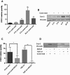
Transforming growth factor beta-1 contributes to a mast cell-induced increase in COX-2 mRNA and protein expression by human tenocytes. Tenocytes were cultured in the presence or absence of HMC-1 conditioned media (cond med), recombinant active transforming growth factor beta-1 (TGF-β1), and A83-01 for the indicated times. Quantitative polymerase chain reaction and western blots show increased cyclooxygenase COX-2 mRNA (A) and protein (B) in response to HMC-1 conditioned media. A83-01 reduced COX-2 mRNA (C) and Cox-2 protein (D) by tenocytes in response to treatment with mast cell conditioned media or TGF-β1 for 6 hours. Data represent mean ± standard error of the mean of at least three independent experiments, each run in triplicate. Mann–Whitney U test. *P = 0.022, **P = 0.0037.
Figure 3
Figure 3
Mast cell-induced prostaglandin E2 production by human tenocytes is mediated through transforming growth factor beta-1. Tenocytes were stimulated with HMC-1 conditioned media (cond media) with or without indomethacin (2 μM) or A83-01 (2 μM) for 6 hours. Media were removed, cells were washed three times with phosphate-buffered saline, and incubated with a fresh serum-free media for 24 hours. Enzyme-linked immunosorbent assay measured the prostaglandin E2 (PGE2) concentration in tenocyte-conditioned media. Data represents mean ± standard error of the mean of at least three separate experiments. One-way analysis of variance, P <0.001. *Statistically significant Bonferroni post-hoc tests of P <0.01.
Figure 4
Figure 4
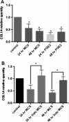
Mast cell-induced modulation of collagen expression by human tenocytes is mediated through prostaglandin E2. Tenocytes cultured in a three-dimensional collagen matrix were stimulated with mast cell sonicate (MCS), prostaglandin E2 (PGE2, 5 μM), or sonicate generated from indomethacin-treated mast cells (Indo MCS). Quantitative polymerase chain reaction analysis shows COL1A1transcription levels by tenocytes in response to MCS and PGE2(A), and MCS versus Indo MCS (B). *P <0.001.
Figure 5
Figure 5
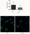
Mast cells reduce type I procollagen protein expression by tenocytes. Immunofluorescent staining for type I procollagen protein in a monolayer of tenocytes stimulated with HMC-1 conditioned media (cond media) for 24 hours. The immunofluorescent images were analyzed for intensity of procollagen immunoreaction using the open source software CellProfiler and data are presented as mean arbitrary fluorescence intensity units (a.u.). Representative immunofluorescent images of control versus conditioned media-treated tenocytes are shown. Data represent mean ± standard error of the mean of at least three separate experiments, each run in triplicate. Mann–Whitney U test, *P <0.001.
Figure 6
Figure 6
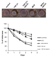
Mast cell-induced contraction of a three-dimensional collagen matrix is mediated through transforming growth factor beta-1. Tenocytes embedded in a three-dimensional collagen gel were stimulated with mast cell sonicate (MCS) or active transforming growth factor beta-1 (TGF-β1), in the presence or absence of A83-01. Data represent mean ± standard error of the mean of at least three separate experiments, each run in triplicate. Two-way analysis of variance followed by Bonferroni’s post-hoc test was used for comparisons. P <0.001 for days 2 to 4 for the following group comparisons: control versus MCS, MCS versus MCS + A83-01, control versus TGF-β1, and TGF-β1 versus TGF-β1 + A83-01.
Figure 7
Figure 7
Matrix metalloproteinases contribute to the mast cell-induced contraction of tenocyte populated collagen matrices. Tenocytes embedded in collagen matrices were stimulated with mast cell sonicate (MCS) for the indicated times and RNA was extracted. The quantitative polymerase chain reaction analysis shows the mRNA expression of matrix metalloproteinases MMP1(A) and MMP7(B) by tenocytes in response to MCS. Data represent mean ± standard error of the mean of at least three separate experiments, each run in triplicate (Student’s t test, *P <0.025 relative to control). (C) A pan-MMP inhibitor (batimastat) added to the collagen gel inhibited contraction of the collagen matrix in response to MCS in a concentration-dependent and time-dependent manner (P <0.001 for effects of both time and treatment).

References

    1. Wygrecka M, Dahal BK, Kosanovic D, Petersen F, Taborski B, von Gerlach S, Didiasova M, Zakrzewicz D, Preissner KT, Schermuly RT, Markart P. Mast cells and fibroblasts work in concert to aggravate pulmonary fibrosis: role of transmembrane SCF and the PAR-2/PKC-alpha/Raf-1/p44/42 signaling pathway. Am J Pathol. 2013;15:2094–2108. doi: 10.1016/j.ajpath.2013.02.013. - DOI - PubMed
    1. Holdsworth SR, Summers SA. Role of mast cells in progressive renal diseases. J Am Soc Nephrol. 2008;15:2254–2261. doi: 10.1681/ASN.2008010015. - DOI - PubMed
    1. Pearson ME, Huff JC, Giorno RC, Panicheewa S, Claman HN, Steigerwald JC. Immunologic dysfunction in scleroderma: evidence for increased mast cell releasability and HLA-DR positivity in the dermis. Arthritis Rheum. 1988;15:672–677. doi: 10.1002/art.1780310514. - DOI - PubMed
    1. Scott A, Lian O, Bahr R, Hart DA, Duronio V, Khan KM. Increased mast cell numbers in human patellar tendinosis: correlation with symptom duration and vascular hyperplasia. Br J Sports Med. 2008;15:753–757. doi: 10.1136/bjsm.2007.040212. - DOI - PubMed
    1. Millar NL, Hueber AJ, Reilly JH, Xu Y, Fazzi UG, Murrell GA, McInnes IB. Inflammation is present in early human tendinopathy. Am J Sports Med. 2010;15:2085–2091. doi: 10.1177/0363546510372613. - DOI - PubMed

Publication types

MeSH terms