

Severity of middle cerebral artery occlusion determines retinal deficits in rats
- PMID: 24518488
- PMCID: PMC4040498
- DOI: 10.1016/j.expneurol.2014.02.005
Severity of middle cerebral artery occlusion determines retinal deficits in rats
Abstract
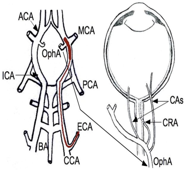
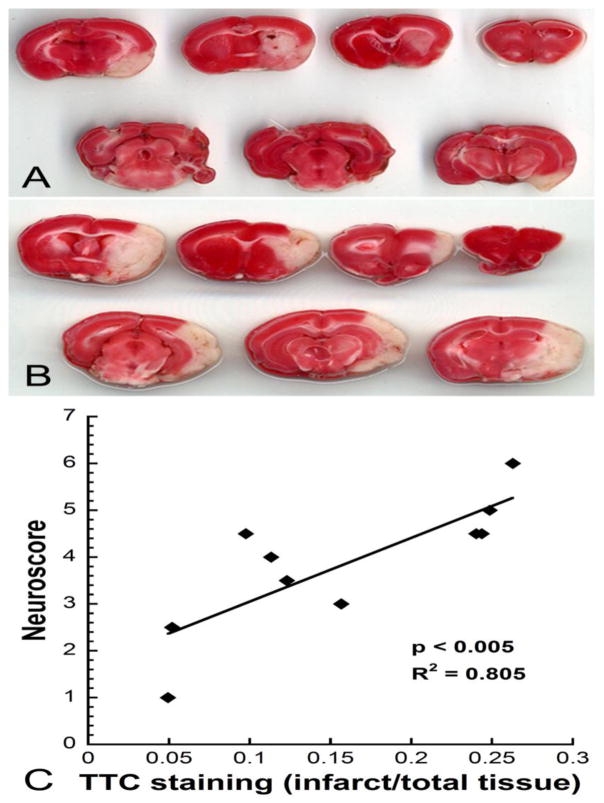
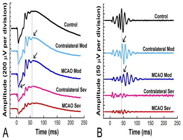
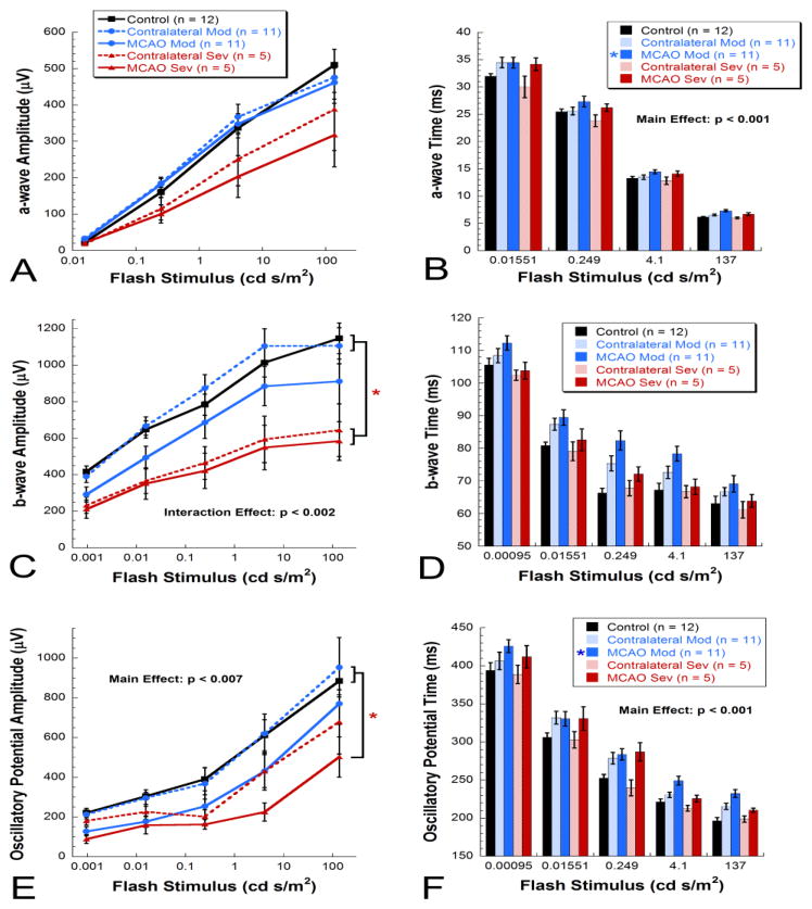
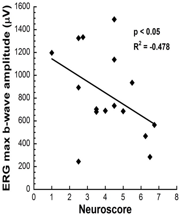
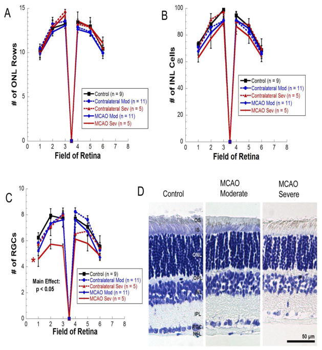
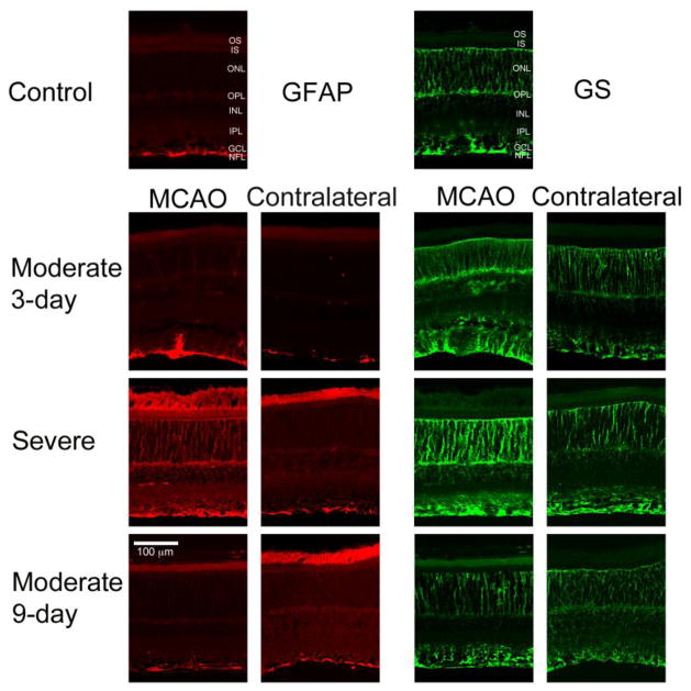
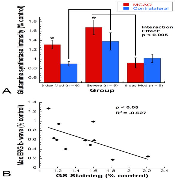
Middle cerebral artery occlusion (MCAO) using the intraluminal suture technique is a common model used to study cerebral ischemia in rodents. Due to the proximity of the ophthalmic artery to the middle cerebral artery, MCAO blocks both arteries, causing both cerebral ischemia and retinal ischemia. While previous studies have shown retinal dysfunction at 48h post-MCAO, we investigated whether these retinal function deficits persist until 9days and whether they correlate with central neurological deficits. Rats received 90min of transient MCAO followed by electroretinography at 2 and 9days to assess retinal function. Retinal damage was assessed with cresyl violet staining, immunohistochemistry for glial fibrillary acidic protein (GFAP) and glutamine synthetase, and TUNEL staining. Rats showed behavioral deficits as assessed with neuroscore that correlated with cerebral infarct size and retinal function at 2days. Two days after surgery, rats with moderate MCAO (neuroscore <5) exhibited delays in electroretinogram implicit time, while rats with severe MCAO (neuroscore ≥5) exhibited reductions in amplitude. Glutamine synthetase was upregulated in Müller cells 3days after MCAO in both severe and moderate animals; however, retinal ganglion cell death was only observed in MCAO retinas from severe animals. By 9days after MCAO, both glutamine synthetase labeling and electroretinograms had returned to normal levels in moderate animals. Early retinal function deficits correlated with behavioral deficits. However, retinal function decreases were transient, and selective retinal cell loss was observed only with severe ischemia, suggesting that the retina is less susceptible to MCAO than the brain. Temporary retinal deficits caused by MCAO are likely due to ischemia-induced increases in extracellular glutamate that impair signal conduction, but resolve by 9days after MCAO.
Keywords: Electroretinogram; Focal ischemia; Middle cerebral artery occlusion; Rat; Retina; Retinal ischemia.
Copyright © 2014 Elsevier Inc. All rights reserved.
Figures
Similar articles
-
Neuroprotective effects of Acer palmatum thumb. leaf extract (KIOM-2015E) against ischemia/reperfusion-induced injury in the rat retina.Mol Vis. 2020 Oct 10;26:691-704. eCollection 2020. Mol Vis. 2020. PMID: 33088173 Free PMC article.
-
Progesterone treatment in two rat models of ocular ischemia.Invest Ophthalmol Vis Sci. 2015 May;56(5):2880-91. doi: 10.1167/iovs.14-16070. Invest Ophthalmol Vis Sci. 2015. PMID: 26024074 Free PMC article.
-
Gonadal steroids block the calpain-1-dependent intrinsic pathway of apoptosis in an experimental rat stroke model.Neurol Res. 2017 Jan;39(1):54-64. doi: 10.1080/01616412.2016.1250459. Epub 2016 Nov 11. Neurol Res. 2017. PMID: 27832728
-
Protective effects of remote ischemic conditioning against ischemia/reperfusion-induced retinal injury in rats.Vis Neurosci. 2014 May;31(3):245-52. doi: 10.1017/S0952523814000121. Epub 2014 Apr 15. Vis Neurosci. 2014. PMID: 24735565
-
Risk of bias reporting in the recent animal focal cerebral ischaemia literature.Clin Sci (Lond). 2017 Oct 12;131(20):2525-2532. doi: 10.1042/CS20160722. Print 2017 Oct 15. Clin Sci (Lond). 2017. PMID: 29026002 Free PMC article. Review.
Cited by
-
Understanding Retinal Changes after Stroke.Open J Ophthalmol. 2017 Nov;7(4):281-292. doi: 10.4236/ojoph.2017.74037. Epub 2017 Nov 6. Open J Ophthalmol. 2017. PMID: 30956896 Free PMC article.
-
Early retinal inflammatory biomarkers in the middle cerebral artery occlusion model of ischemic stroke.Mol Vis. 2016 Jun 4;22:575-88. eCollection 2016. Mol Vis. 2016. PMID: 27293375 Free PMC article.
-
NLRP3 Inflammasome: A Potential Target in Isoflurane Pretreatment Alleviates Stroke-Induced Retinal Injury in Diabetes.Front Cell Neurosci. 2021 Jul 8;15:697449. doi: 10.3389/fncel.2021.697449. eCollection 2021. Front Cell Neurosci. 2021. PMID: 34305534 Free PMC article.
-
Redefining the Koizumi model of mouse cerebral ischemia: A comparative longitudinal study of cerebral and retinal ischemia in the Koizumi and Longa middle cerebral artery occlusion models.J Cereb Blood Flow Metab. 2022 Nov;42(11):2080-2094. doi: 10.1177/0271678X221109873. Epub 2022 Jun 24. J Cereb Blood Flow Metab. 2022. PMID: 35748043 Free PMC article.
-
Anti-inflammatory α-Melanocyte-Stimulating Hormone Protects Retina After Ischemia/Reperfusion Injury in Type I Diabetes.Front Neurosci. 2022 Feb 25;16:799739. doi: 10.3389/fnins.2022.799739. eCollection 2022. Front Neurosci. 2022. PMID: 35281489 Free PMC article.
References
-
- Acar N, Bonhomme B, Joffre C, Bron AM, Creuzot-Garcher C, Bretillon L, Doly M, Chardigny JM. The retina is more susceptible than the brain and the liver to the incorporation of trans isomers of DHA in rats consuming trans isomers of alpha-linolenic acid. Reproduction, nutrition, development. 2006;46:515–525. - PubMed
-
- Andersen MB, Zimmer J, Sams-Dodd F. Specific behavioral effects related to age and cerebral ischemia in rats. Pharmacology, biochemistry, and behavior. 1999;62:673–682. - PubMed
-
- Barnett NL, Pow DV. Antisense knockdown of GLAST, a glial glutamate transporter, compromises retinal function. Investigative ophthalmology & visual science. 2000;41:585–591. - PubMed
-
- Beal MF. Mechanisms of excitotoxicity in neurologic diseases. FASEB journal: official publication of the Federation of American Societies for Experimental Biology. 1992;6:3338–3344. - PubMed
-
- Benavente O, Eliasziw M, Streifler JY, Fox AJ, Barnett HJ, Meldrum H. Prognosis after transient monocular blindness associated with carotid-artery stenosis. The New England journal of medicine. 2001;345:1084–1090. - PubMed
Publication types
MeSH terms
Substances
Grants and funding
LinkOut - more resources
Full Text Sources
Other Literature Sources
Medical
Miscellaneous
