Review
. 2014;8(3):269-86.
doi: 10.1007/s12105-014-0527-7. Epub 2014 Feb 12.

Carcinoma ex-Schneiderian papilloma (malignant transformation): a clinicopathologic and immunophenotypic study of 20 cases combined with a comprehensive review of the literature

Affiliations
Review

Carcinoma ex-Schneiderian papilloma (malignant transformation): a clinicopathologic and immunophenotypic study of 20 cases combined with a comprehensive review of the literature

Jeremy Nudell et al. Head Neck Pathol. 2014.

Abstract

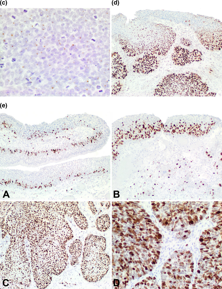
Schneiderian papilloma (SP) are uncommon tumors with malignant transformation even less common. The histologic criteria to define malignant transformation are not well developed nor is the immunohistochemical profile reported in a large series of carcinomas. 20 cases of malignant transformation of SP included 7 females and 13 males, aged 38-86 years (mean 60.7 years). Patients presented most frequently with a mass (n = 11) and obstructive symptoms (n = 7), present for 38.7 months (mean). Most patients had no previous history of SP (n = 13); metachronous carcinoma was identified in 7 patients an average of 34.4 months after the first diagnosis of SP, with 1-4 recurrences of SP. With a mean size of 4.1 cm, the majority of tumors involved a combination of more than one anatomic site (n = 10), followed by the maxillary sinus only (n = 5) or nasal cavity only (n = 3). Histologically, 17 were inverted and 3 exophytic type SP. There were 17 squamous cell carcinomas, 2 mucoepidermoid carcinomas and 1 sinonasal undifferentiated carcinoma, comprising from 10 to 95 % of the tumor volume. Malignant histologic features included atypical mitoses, necrosis, bone invasion, lymphovascular invasion, decreased transmigrating neutrophils, paradoxical maturation, dyskeratosis and/or perineural invasion (n = 3). Patients tended to present with advanced stage (n = 14, Stage III and IV). Immunohistochemical studies showed positive reactions in the malignancies for CK5/6 (86 %), p63 (86 %), CK7 (luminal, 50 %), p53 (83 %), and p16 (25 %). In situ hybridization detected human papillomavirus in 26 %. Surgery was often accompanied by radiation therapy (n = 13), with a mean of 2.4 years of follow-up. Five patients developed a recurrence between 0.8 and 3.3 years. Carcinomas ex-SP are less common and are associated with better outcome than previously reported. Patients tend to present with a synchronous carcinoma, developing in an inverted type SP, with squamous cell carcinoma the most common malignancy. Development of metachronous carcinomas ex-SP was always preceded by SP recurrence in this series.


Figures

Fig. 1
Fig. 1
a This composite image demonstrates several zones of transition. A The left shows ciliated respiratory epithelium that abruptly transitions to carcinoma in situ. B The left shows a Schneiderian papilloma that abruptly transitions into an area of carcinoma. C This inverted papilloma contains areas of Pagetoid spread of the carcinoma (SNUC was noted below). D Paradoxical maturation is noted at the base of this dysplastic area, with a small area suggestive of early invasion. b This composite highlights areas of transition between benign and malignant. A The left portion shows a malignant transformation that blends with the right sided benign component. B Three different fields show unremarkable ciliated epithelium, dysplasia, and carcinoma in situ. C The upper portion of the field shows a Schneiderian papilloma, while the lower field shows malignant transformation. D The upper field demonstrates koilocytic atypia of a benign papilloma, while the lower field shows full thickness carcinoma in situ
Fig. 2
Fig. 2
a This mucoepidermoid carcinoma shows an epidermoid and transitional epithelium, with mucocytes within the tumor. There are no cilia to suggest they may be surface epithelium. b A non-keratinizing squamous cell carcinoma showing lymphoepithelial-like features in this poorly differentiated non-keratinizing squamous cell carcinoma. c A sinonasal undifferentiated carcinoma was the malignancy noted in one case
Fig. 3
Fig. 3
a A composite of various cytologic features in malignant transformation. A Koilocytic atypia, pleomorphism and architectural disorganization. B Focal nuclear molding with cells that have a high nuclear to cytoplasmic ratio. Cell borders are prominent. C Increased mitoses (15 in this field), including atypical forms (off center), seen in carcinoma. D Dyskeratosis and keratin pearl formation. b A A benign Schneiderian papilloma with well developed transepithelial migration of neutrophils. B Neutrophils were greatly reduced or absent in areas of carcinoma. c A A complex inverted and destructive architecture, but the spicules of bone are native, and not destroyed. B The spicule of bone is being destroyed by highly atypical squamous epithelium, associated with a desmoplastic stroma. Note osteoblasts and osteoclasts. C Vascular invasion by a tumor thrombus. d A Central comedonecrosis with keratin debris is noted in this area of carcinoma. B Ghost cell outlines along with degenerated and necrotic debris are intermixed with the viable neoplastic cells in this area of necrosis
Fig. 3
Fig. 3
a A composite of various cytologic features in malignant transformation. A Koilocytic atypia, pleomorphism and architectural disorganization. B Focal nuclear molding with cells that have a high nuclear to cytoplasmic ratio. Cell borders are prominent. C Increased mitoses (15 in this field), including atypical forms (off center), seen in carcinoma. D Dyskeratosis and keratin pearl formation. b A A benign Schneiderian papilloma with well developed transepithelial migration of neutrophils. B Neutrophils were greatly reduced or absent in areas of carcinoma. c A A complex inverted and destructive architecture, but the spicules of bone are native, and not destroyed. B The spicule of bone is being destroyed by highly atypical squamous epithelium, associated with a desmoplastic stroma. Note osteoblasts and osteoclasts. C Vascular invasion by a tumor thrombus. d A Central comedonecrosis with keratin debris is noted in this area of carcinoma. B Ghost cell outlines along with degenerated and necrotic debris are intermixed with the viable neoplastic cells in this area of necrosis
Fig. 4
Fig. 4
a A composite image of p16 immunoreactivity. A Strong, diffuse, nuclear and cytoplasmic reaction in the benign and malignant areas of this case. B Strong nuclear reaction (left sided) in an area of malignant transformation, mixed nuclear and cytoplasmic reactivity in the same area, while the area of benign papilloma showed a different pattern (lower). C Strong, diffuse, nuclear reaction of the whole epithelium in an area of carcinoma. D No reaction in an area of carcinoma, but the stroma shows a strong and diffuse nuclear and cytoplasmic reaction in the fibroblastic cells. b There was quite variable reactivity with p16. A: In an area of early invasion, there is nuclear and cytoplasmic strong reactivity, while the remaining area is negative. B This carcinoma showed strong and diffuse nuclear and cytoplasmic reactivity. c A delicate, single nuclear dot reaction with HPV ISH in an area of carcinoma. d p53 showed a gradient of reactivity, with a greater percentage of the nuclei showing a positive reaction in areas of carcinoma compared to benign areas. There was a greater intensity of the stain in areas of malignancy. e Ki-67 immunoreactivity. A: Benign Schneiderian papilloma with a limited, basal reaction. B Increased number of nuclei positive and above the basal zone in an area of severe dysplasia. C Most of the nuclei are positive in this area of carcinoma. D Very strong, heavy nuclear reaction was present in areas of carcinoma
Fig. 4
Fig. 4
a A composite image of p16 immunoreactivity. A Strong, diffuse, nuclear and cytoplasmic reaction in the benign and malignant areas of this case. B Strong nuclear reaction (left sided) in an area of malignant transformation, mixed nuclear and cytoplasmic reactivity in the same area, while the area of benign papilloma showed a different pattern (lower). C Strong, diffuse, nuclear reaction of the whole epithelium in an area of carcinoma. D No reaction in an area of carcinoma, but the stroma shows a strong and diffuse nuclear and cytoplasmic reaction in the fibroblastic cells. b There was quite variable reactivity with p16. A: In an area of early invasion, there is nuclear and cytoplasmic strong reactivity, while the remaining area is negative. B This carcinoma showed strong and diffuse nuclear and cytoplasmic reactivity. c A delicate, single nuclear dot reaction with HPV ISH in an area of carcinoma. d p53 showed a gradient of reactivity, with a greater percentage of the nuclei showing a positive reaction in areas of carcinoma compared to benign areas. There was a greater intensity of the stain in areas of malignancy. e Ki-67 immunoreactivity. A: Benign Schneiderian papilloma with a limited, basal reaction. B Increased number of nuclei positive and above the basal zone in an area of severe dysplasia. C Most of the nuclei are positive in this area of carcinoma. D Very strong, heavy nuclear reaction was present in areas of carcinoma

References

    1. AJCC Cancer Staging Manual, 7th ed. New York: Springer; 2009.
    1. Buchwald C, Franzmann MB, Jacobsen GK, Juhl BR, Lindeberg H. Carcinomas occurring in papillomas of the nasal septum associated with human papilloma virus (HPV) Rhinology. 1997;35:74–78. - PubMed
    1. Dingle I, Stachiw N, Bartlett A, Lambert P. Bilateral inverted papilloma of the middle ear with intracranial involvement and malignant transformation: first reported case. Laryngoscope. 2012;122:1615–1619. doi: 10.1002/lary.23247. - DOI - PubMed
    1. Dolgin SR, Zaveri VD, Casiano RR, Maniglia AJ. Different options for treatment of inverting papilloma of the nose and paranasal sinuses: a report of 41 cases. Laryngoscope. 1992;102:231–236. doi: 10.1288/00005537-199203000-00001. - DOI - PubMed
    1. Halimi M, Aghbali A, Emamverdizadeh P, Talesh KT. Inverted papilloma of the palate with malignant transformation. J Oral Maxillofac Pathol. 2012;16:291–293. doi: 10.4103/0973-029X.99093. - DOI - PMC - PubMed

Substances

LinkOut - more resources