. 2014 Apr;5(2):253-79.
doi: 10.1007/s13244-014-0309-5. Epub 2014 Feb 12.

Ultrasonography for clinical decision-making and intervention in airway management: from the mouth to the lungs and pleurae

Affiliations

Ultrasonography for clinical decision-making and intervention in airway management: from the mouth to the lungs and pleurae

Michael S Kristensen et al. Insights Imaging. 2014 Apr.

Abstract

Objectives: To create a state-of-the-art overview of the new and expanding role of ultrasonography in clinical decision-making, intervention and management of the upper and lower airways, that is clinically relevant, up-to-date and practically useful for clinicians.

Methods: This is a narrative review combined with a structured Medline literature search.

Results: Ultrasonography can be utilised to predict airway difficulty during induction of anaesthesia, evaluate if the stomach is empty or possesses gastric content that poses an aspiration risk, localise the essential cricothyroid membrane prior to difficult airway management, perform nerve blocks for awake intubation, confirm tracheal or oesophageal intubation and facilitate localisation of tracheal rings for tracheostomy. Ultrasonography is an excellent diagnostic tool in intraoperative and emergency diagnosis of pneumothorax. It also enables diagnosis and treatment of interstitial syndrome, lung consolidation, atelectasis, pleural effusion and differentiates causes of acute breathlessness during pregnancy. Patient safety can be enhanced by performing procedures under ultrasound guidance, e.g. thoracocentesis, vascular line access and help guide timing of removal of chest tubes by quantification of residual pneumothorax size.

Conclusions: Ultrasonography used in conjunction with hands-on management of the upper and lower airways has multiple advantages. There is a rapidly growing body of evidence showing its benefits.

Teaching points: • Ultrasonography is becoming essential in management of the upper and lower airways. • The tracheal structures can be identified by ultrasonography, even when unidentifiable by palpation. • Ultrasonography is the primary diagnostic approach in suspicion of intraoperative pneumothorax. • Point-of-care ultrasonography of the airways has a steep learning curve. • Lung ultrasonography allows treatment of interstitial syndrome, consolidation, atelectasis and effusion.


Figures

Fig. 1
Fig. 1
Mouth and tongue. Left: The curved, low frequency transducer and the area covered by the scanning (light blue). Middle: The resulting ultrasound image. Right: The dorsal surface of the tongue (orange), the muscles in the floor of the mouth (turquoise). The shadows (brown) from the mentum of the mandible anteriorly and from the hyoid bone posteriorly
Fig. 2
Fig. 2
Midline sagittal scan from the hyoid bone to the proximal part of the thyroid cartilage. Left: The black outline shows the area covered by the scanning. Middle: The scanning image. Right: The shadow from the hyoid bone (brown); the thyro-hyoid membrane (yellow); posterior surface of part of epiglottis (red); pre-epiglottic fat (green); thyroid cartilage (purple)
Fig. 3
Fig. 3
Larynx and vocal cords. Left: Transverse midline scan over the thyroid cartilage (in an 8-year-old boy). Vocal cords (dark blue); anterior commisure (light blue); arytenoid cartilages (green); thyroid cartilage (yellow)
Fig. 4
Fig. 4
Cricothyroid membrane. Left: The linear high frequency transducer placed in the midsagittal plane, the scanning area is marked with a black line. Right: The cricothyroid membrane (orange); the thyroid cartilage (green); the cricoid cartilage (purple); anterior part of tracheal rings (dark blue); the tissue/air border (light blue); the isthmus of the thyroid gland (yellow). Below the tissue/air border only artefacts are seen (white)
Fig. 5
Fig. 5
Oesophagus and trachea. Transverse scan just cranial to the suprasternal notch and to the patient’s left side of the trachea. Anterior part of tracheal cartilage (dark blue); oesophagus (yellow); carotid artery (red). Below the tissue/air border in the trachea only artefacts are seen (white)
Fig. 6
Fig. 6
Scanning protocol used for a focused LUS examination. The anterior and posterior axillary line can be used to divide each hemithorax into an anterior, lateral and posterior surface. a The anterior and lateral surfaces on each side of the chest are both divided into an upper and lower quadrant, the image demonstrates the four quadrants on the left hemithorax (1L–4L). b The posterior surfaces on each side were divided into an upper, middle and lower quadrant, the image demonstrates the four quadrants on the left hemithorax (5L–7L). Each quadrant represents a scanning zone, in which the transducer is placed approximately in the middle of the zone making a cross sectional image of an intercostal space and the underlying pleura blades
Fig. 7
Fig. 7
Normal LUS findings: a The transducer is placed in a longitudinal axis over an intercostal space at the anterior surface of the chest. b In the corresponding B-mode image, two ribs are visible aligning the intercostal space, as two hyperechoic lines with an underlying shadow. Lying deeper between the two ribs, a hyperechoic horizontal line is seen representing the visceral and parietal pleura. Using B-mode the movement of the pleural line is called “lung sliding”. c If M-mode scanning is applied, a characteristic pattern called “seashore sign” is visible. The pleural line appears as a hyperechoic line, with the more superficially placed structures appearing as horizontal lines similar to the sea and the part of the picture below the pleural line is grittier looking, mimicking the sand on a seashore
Fig. 8
Fig. 8
Stomach imaging. Left: Probe position—this shows a sagittal probe orientation in the epigastric area. Right: The antrum (Ant) located immediately posterior to the left lobe of the liver (L). The pancreas (P) is typically hyperechoic and located posterior to the antrum. In this figure a cross-section of the splenic vein (SV) may be seen as it crosses the pancreas from right to left. Posterior to the pancreas you can see a longitudinal view of the aorta (Ao). Spine (S). (Courtesy of Anahi Perlas, University of Toronto and Toronto Western Hospital, Toronto, Canada)
Fig. 9
Fig. 9
Hyomental distance ratio. The hyomental distance measured with the head in neutral position (left) and in maximum extension (right). In this case the ratio was only 1.01 and the patient correspondingly had a difficult intubation (Cormack-Lehane grade 4). (Courtesy of Jacek A. Wojtczak, Department of Anesthesiology, University of Rochester School of Medicine and Dentistry, Rochester, NY, USA)
Fig. 10
Fig. 10
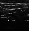
Zenker diverticulum. Transverse scan on the anterior neck above the suprasternal notch shoving a Zenker diverticulum lateral to the trachea. A bolus of solid food is seen in the diverticulum. Sternocleid sternocleidomastoid muscle; Carotid common carotid artery. (Courtesy of Peter Cheng, Kaiser Permanente Riverside Medical Center, Riverside, CA, USA)
Fig. 11
Fig. 11
The stomach. Left Empty state (low aspiration risk). The empty antrum (Ant) appears as a small round or oval structure. It may resemble a “bull’s eye target”. When the antrum is empty, all you can see is gastric wall. What appears to be a small amount of content is actually the thickness of all the layers of the gastric wall. The gastric wall has five distinct sonographic layers. The most prominent layer can be clearly seen in this figure as a hypoechoic “ring” and it corresponds histologically to the muscularis propriae of the stomach. The antrum is located immediately posterior to the left lobe of the liver (L); the pancreas (P); splenic vein (SV); aorta (Ao); spine (S). Middle: Solid content (high aspiration risk) Solid content in the stomach appears as non-homogeneous, mostly hyperechoic content. There is usually some amount of air mixed with the solid meal, and this produces multiple “ring down artefacts” that obscure the posterior wall. We call this type of image a “frosted glass pattern”, and we can see some degree of this type of artefact in this image. Right Clear fluid content. Clear fluid in the stomach (such as water, tea or normal gastric secretions) can be seen as a homogeneous hypoechoic content within the antrum. When clear fluid is seen, it may be useful to do a volume estimation to better assess aspiration risk. (Courtesy of Anahi Perlas, University of Toronto and Toronto Western Hospital, Toronto, Canada)
Fig. 12
Fig. 12
Localisation of the displaced trachea. The patient previously had surgery and radiation therapy for neck cancer and no structures could be palpated. Left: The transducer is placed transversely in the midline over the suprasternal notch. Middle: The scanning image. Right: The cartilage of the tracheal ring (dark blue) is deviated to the patient’s left side. The green line indicates the midline of the neck
Fig. 13
Fig. 13
Localisation of the cricothyroid membrane. Orange is the anterior part of the tracheal rings. Red is the anterior part of the cricoid cartilage. The turquoise line is the shadow from the needle that has been slid underneath the transducer and placed just cranial to the cricoid cartilage, thus indicating the position of the lower part of the cricothyroid membrane, where an emergency airway access should be performed. a The patient is lying supine and operator stands on the patient’s right side facing the patient. b The linear transducer is placed transversely over the neck just above the suprasternal notch and the trachea is observed in the midline. c The transducer is moved to the patient’s right side so that the right border of the transducer is superficial to the midline of the trachea. d The right end of the transducer is kept in the midline of the trachea while the left end of the transducer is rotated into the sagittal plane, resulting in a longitudinal scan of the midline of the trachea; the caudal part of the cricoid cartilage is seen (red). e The transducer is moved cranially and the cricoid cartilage (red) is seen as a slightly elongated structure that is significantly larger and more anteriorly than the tracheal rings. f A needle is moved under the transducer from the cranial end, used only as a marker. The shadow (turquoise line) is just cranial to the cranial border of the cricoid cartilage. g The transducer is removed and the needle indicates the distal part of the cricothyroid membrane
Fig. 14
Fig. 14
Pneumothorax. a The transducer is placed in a longitudinal axis over an intercostal space at the anterior surface of the chest; this is the area where free air in the chest cavity is expected to be located. b If pneumothorax is present the pleura-line only represents the parietal pleura, therefore the motion of the visceral pleura cannot be visualised and subsequently lung sliding is absent. c In M-mode the lack of lung sliding will appear as the “stratosphere sign”, a pattern only consisting of horizontal lines
Fig. 15
Fig. 15
Algorithm for the diagnosis and exclusion of pneumothorax using LUS. Initially one should look for signs which rule out the presence of pneumothorax [lung sliding, lung pulse, B-line(s)] at the anterior surface of the chest. If none of these are present, then one should gradually move the transducer laterally and posterior on the surface of the chest and look for lung point in order to establish the diagnosis of pneumothorax. If neither signs are present, contemplation is needed since pneumothorax can neither be ruled in nor out. In young, previously healthy patients, such as most trauma patients, the absence of lung sliding alone is sufficient to diagnose pneumothorax. In such patients the absence of all signs will be consistent with pneumothorax. In comparison, patients with known lung diseases or previous chest surgery may have a variety of causes for the absence of lung sliding. In such patients the absence of all signs can neither be used to rule in nor to rule out a pneumothorax and further imaging should be performed in order to establish whether pneumothorax is present or absent
Fig. 16
Fig. 16
Multiple B-lines. a The transducer is placed in a longitudinal axis over an intercostal space. b B-lines (arrowheads) are seen as strong hyperechoic, laser-like, vertical lines originating from the pleura and extending to the bottom of the field of view without decreasing in intensity. B-lines move synchronously with lung sliding
Fig. 17
Fig. 17
a Consolidation: A diffusely demarcated consolidation (Pnm) with an appearance similar to liver tissue. Air bronchograms (arrowhead) are visible within the consolidation. b Lung consolidation due to pulmonary embolism. A sharply defined hypoechogenic consolidation (PE) without air bronchograms just below the pleural line. c Tumour. Just below a pleural effusion (Ef), a relatively well demarcated tumour (T) can be seen located on the surface and within the lung tissue. d Lung contusion. US image from a patient who had suffered blunt trauma to the left side of the chest. LUS revealed multiple rib fractures and an underlying lung contusion (Lc) just cranially to the spleen (Spl)
Fig. 18
Fig. 18
Pleural effusion. a The transducer is placed in a longitudinal axis over an intercostal space on the most dependent area of the chest, since this is the area where free fluid in the chest cavity is expected to be located. b Pleural effusion (Eff) often is visualised as a dark, anechoic area lying in between the visceral and parietal pleura on ultrasonography and just above the liver (Lvr) or spleen. Underlying compression atelectasis (Atl) of the lung is also often present

References

    1. Cook TM, Woodall N, Harper J, Benger J. Major complications of airway management in the UK: results of the fourth national audit project of the royal college of anaesthetists and the difficult airway society. Part 2: intensive care and emergency departments. Br J Anaesth. 2011;106:632–642. - PubMed
    1. Lichtenstein D, Meziere G, Biderman P, Gepner A, Barre O. The comet-tail artifact. An ultrasound sign of alveolar-interstitial syndrome. Am J Respir Crit Care Med. 1997;156:1640–1646. - PubMed
    1. Baldi G, Gargani L, Abramo A, et al. Lung water assessment by lung ultrasonography in intensive care: a pilot study. Intensive Care Med. 2013;39:74–84. - PubMed
    1. Soldati G, Inchingolo R, Smargiassi A, et al. Ex vivo lung sonography: morphologic-ultrasound relationship. Ultrasound Med Biol. 2012;38:1169–1179. - PubMed
    1. Via G, Lichtenstein D, Mojoli F, et al. Whole lung lavage: a unique model for ultrasound assessment of lung aeration changes. Intensive Care Med. 2010;36:999–1007. - PubMed