. 2014 May;23(5):603-17.
doi: 10.1002/pro.2442. Epub 2014 Mar 19.

Influence of hydrophobic and electrostatic residues on SARS-coronavirus S2 protein stability: insights into mechanisms of general viral fusion and inhibitor design

Affiliations

Influence of hydrophobic and electrostatic residues on SARS-coronavirus S2 protein stability: insights into mechanisms of general viral fusion and inhibitor design

Halil Aydin et al. Protein Sci. 2014 May.

Abstract

Severe acute respiratory syndrome (SARS) is an acute respiratory disease caused by the SARS-coronavirus (SARS-CoV). SARS-CoV entry is facilitated by the spike protein (S), which consists of an N-terminal domain (S1) responsible for cellular attachment and a C-terminal domain (S2) that mediates viral and host cell membrane fusion. The SARS-CoV S2 is a potential drug target, as peptidomimetics against S2 act as potent fusion inhibitors. In this study, site-directed mutagenesis and thermal stability experiments on electrostatic, hydrophobic, and polar residues to dissect their roles in stabilizing the S2 postfusion conformation was performed. It was shown that unlike the pH-independent retroviral fusion proteins, SARS-CoV S2 is stable over a wide pH range, supporting its ability to fuse at both the plasma membrane and endosome. A comprehensive SARS-CoV S2 analysis showed that specific hydrophobic positions at the C-terminal end of the HR2, rather than electrostatics are critical for fusion protein stabilization. Disruption of the conserved C-terminal hydrophobic residues destabilized the fusion core and reduced the melting temperature by 30°C. The importance of the C-terminal hydrophobic residues led us to identify a 42-residue substructure on the central core that is structurally conserved in all existing CoV S2 fusion proteins (root mean squared deviation=0.4 Å). This is the first study to identify such a conserved substructure and likely represents a common foundation to facilitate viral fusion. We have discussed the role of key residues in the design of fusion inhibitors and the potential of the substructure as a general target for the development of novel therapeutics against CoV infections.

Keywords: MERS-CoV; S2; SARS-CoV; coronavirus; glycoprotein; viral entry; viral fusion.


Figures

Figure 1
Figure 1
Structural description and biophysical characterization of the SARS-CoV S2 L2H protein. (A) Schematic diagram of the SARS-CoV S protein. The S protein exhibits the characteristic domain organization of class I viral proteins. Abbreviations are as follows: S1, CoV attachment subunit; S2, CoV fusion subunit; SP, signal peptide; RBD, receptor binding domain; RBM, receptor binding motif; FP, fusion peptide; HR1, heptad repeat 1 region; HR2, heptad repeat 2 region; T, tether region; TM, transmembrane domain; CT, cytoplasmic tail; L2H, linked two-heptad construct. The positions of the S1 domain (residues 14–667), S2 domain (residues 668–1255), SP (residues 1–14), RBD (residues 306–527), RBM (residues 424–494), HR1 (residues 890–973), tether, and HR2 (residues 1142–1184), TM and CT (residues 1196–1255) are shown above the schematic. Red arrows indicate the S1–S2 and S′ proteolytic cleavage sites at residues R667 and R797, respectively. SARS-CoV S2 L2H construct was generated by using HR1 residues 896–972 and tether/HR2 residues 1142–1183 connected by a six amino acid linker at the HR1 C-terminal and HR2 N-terminal ends (colored in orange). (B) Sedimentation equilibrium data for a 20 μM sample at 4°C and 22,000 rpm in TBS buffer. The curve indicates the distribution of a 48.4-kDa protein. The data fit closely to a trimeric model for SARS-CoV S2 L2H. The deviation in the data from the linear fit for a trimeric model is plotted in the upper panel. (C) Experimental CD wavelength scan of SARS-CoV S2 L2H (blue) at 25°C reveals minimas at 208 and 222 nm, indicative of strong α-helical secondary structural characteristics. The SARS-CoV S2 L2H is calculated to contain 50% α-helical content. A reconstructed CD wavelength scan (red) shows the quality of the fit used in the calculation of secondary structural content.
Figure 2
Figure 2
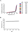
SARS-CoV S2 L2H stability at various pH values. (A) Thermal denaturation of SARS-CoV S2 L2H monitored by CD molar ellipticity at 222 nm in sodium acetate buffer (between pH 4.0 and 6.5), and TBS buffer (between pH 7.0 and 8.5). The CD signal was baseline corrected, normalized between 0 (folded) and 1 (unfolded), and fit to a non-linear biphasic sigmoidal curve. The Tm values correspond to the temperature where 50% of the protein has unfolded. (B) Plot of SARS-CoV S2 L2H stability as a function of pH. The trimeric SARS-CoV S2 L2H is stable between pH values 4.0 and 8.5.
Figure 3
Figure 3
Biophysical characterization of SARS-CoV S2 electrostatic interactions. (A) Ribbon diagram of SARS-CoV S2 fusion core (PDB code: 2BEZ) shows electrostatic interactions between the HR1–HR1 and HR1–HR2 regions. The HR1 and tether/HR2 regions are depicted in gray and green, respectively. The side chains of the ion-pair interactions are colored in magenta. The zoomed views of ion-pairs are shown in the inset boxes and the distances between the residues are indicated in Ångstroms (Å). (B) Thermal denaturation profiles of wild-type (WT), single, (C) double and triple mutant of electrostatic residues in the SARS-CoV S2 fusion subunit. Thermal stability was recorded at 222 nm. All data were baseline corrected, normalized between 0 (folded) and 1 (unfolded) and plotted as a function of temperature. The Tm values indicate the midpoint melting temperatures for WT and mutant proteins.
Figure 4
Figure 4
SARS-CoV S2 hydrophobic and polar interactions. (A) Ribbon diagram of SARS-CoV S2 fusion core structure (PDB Code: 2BEZ). The HR1 region forms a long helical strand with 22 helical turns (colored in gray). The tether and HR2 regions extend alongside the HR1 inner core in an antiparallel manner (colored in green). The hydrophobic residues at the HR1–tether and HR1–HR2 interface are depicted in orange. The inset boxes show the zoomed view of critical hydrophobic residues positioned at the interfaces. (B) Thermal denaturation of wild-type (WT) and mutant hydrophobic residues in the SARS-CoV fusion subunit. (C) Ribbon diagram of an extended SARS-CoV S2 fusion core structure (PDB Code: 1WYY) displaying two putative chloride binding sites. Chloride ions observed in the crystal structure of the HR1 inner core are shown in red. The polar residues interacting with chloride ion (Q902 and N937) and a single polar residue (N951) at the HR1-tether interface are shown as blue sticks. The HR1 and tether/HR2 regions are depicted in gray and green, respectively. (D) Thermal denaturation profiles of wild-type (WT) and chloride binding site mutants. All thermal denaturation profiles are plotted as described in Figure 3.
Figure 5
Figure 5
Primary sequence alignment of CoV S2 fusion cores. Multiple sequence alignment of various human and animal CoV fusion proteins. Abbreviations are as follows: SARS-CoV, severe acute respiratory syndrome-coronavirus; MERS-CoV, middle east respiratory syndrome-coronavirus; hCoV, human coronavirus; HKU, Hong Kong University strain; BCoV, bovine coronavirus; BtCoV, bat coronavirus; CCoV, canine coronavirus; FCoV, feline coronavirus; FIPV, feline infectious peritonitis virus; MHV, mouse hepatitis virus; MuCoV, munia coronavirus; PEDV, porcine epidemic bronchitis virus; PRCoV, porcine respiratory coronavirus; PHEV, porcine hemagglutinating encephalomyelitis virus; TGEV, transmissible gastroenteritis virus; RbCoV, rabbit coronavirus; RtCoV, rat coronavirus; SpCoV, sparrow coronavirus; ThCoV, thrush coronavirus. Sequence boundaries for HR1 and tether/HR2 regions are depicted with gray and green lines, respectively. Residue numbers corresponding to the SARS-CoV S2 fusion subunit numbering are indicated above the alignment. Strictly conserved residues are outlined in red and residues that are important for the stability of the SARS-CoV S2 fusion core are highlighted in yellow and marked with an asterisk (*). Residues (911–924) involved in the formation of the common HR1 substructure are shown in a black box. The heptad repeat register (a, b, c, d, e, f, g) of the SARS-CoV S2 fusion core is indicated below the alignment.
Figure 6
Figure 6
The common HR1 SARS-CoV S2 substructure. (A) Ribbon diagram of the structural alignments of the HR1 substructure. The SARS-CoV HR1 substructure (colored in green) was superimposed with MHV (colored in purple), MERS-CoV (colored in magenta), and hCoV-NL63 (colored in orange) HR1 substructures (RMSD = ∼0.4 Å). Conserved side chains within the HR1 substructure are shown for each virus. SARS-CoV S2 residue numbering was used in both structural alignments. (B) Characterization of the groove on the surface of the HR1 inner core. The electrostatic surface potential of the HR1 region at the HR2 interface was depicted for the SARS-CoV S2, MHV S2, MERS-CoV S2, and hCoV-NL63 S2 fusion cores. HR2 helical region residues extending alongside the HR1 inner core are shown as yellow sticks. Conserved HR2 residues interacting with the HR1 inner core are labeled accordingly. The structurally conserved HR1 core boundaries are indicated between the black dashed lines. Using computational solvent mapping, a hydrophobic pocket was identified, as shown by the green small molecules, on the HR1 substructure.

References

    1. Perlman S, Netland J. Coronaviruses post-SARS: update on replication and pathogenesis. Nat Rev Microbiol. 2009;7:439–450. - PMC - PubMed
    1. Ksiazek TG, Erdman D, Goldsmith CS, Zaki SR, Peret T, Emery S, Tong S, Urbani C, Comer JA, Lim W, Rollin PE, Dowell SF, Ling A, Humphrey CD, Shieh W, Guarner J, Paddock CD, Rota P, Fields B, DeRisi J, Yang J, Cox N, Hughes JM, LeDuc JW, Bellini WJ, Anderson LJ. A novel coronavirus associated with severe acute respiratory syndrome. N Engl J Med. 2003;348:1953–1966. and the SARS Working Group. - PubMed
    1. Rota PA, Oberste MS, Monroe SS, Nix WA, Campagnoli R, Icenogle JP, Peñaranda S, Bankamp B, Maher K, Chen M, Tong S, Tamin A, Lowe L, Frace M, DeRisi JL, Chen Q, Wang D, Erdman DD, Peret TCT, Burns C, Ksiazek TG, Rollin PE, Sanchez A, Liffick S, Holloway B, Limor J, McCaustland K, Olsen-Rasmussen M, Fouchier R, Günther S, Osterhaus ADME, Drosten C, Pallansch MA, Anderson LJ, Bellini WJ. Characterization of a novel coronavirus associated with severe acute respiratory syndrome. Science. 2003;300:1394–1399. - PubMed
    1. Drosten C, Günther S, Preiser W, van der Werf S, Brodt H, Becker S, Rabenau H, Panning M, Kolesnikova L, Fouchier RAM, Berger A, Burguière A, Cinatl J, Eickmann M, Escriou N, Grywna K, Kramme S, Manuguerra J, Müller S, Rickerts V, Stürmer M, Vieth S, Klenk H, Osterhaus ADME, Schmitz H, Doerr HW. Identification of a novel coronavirus in patients with severe acute respiratory syndrome. N Engl J Med. 2003;348:1967–1976. - PubMed
    1. Gerlier D. Emerging zoonotic viruses: new lessons on receptor and entry mechanisms. Curr Opin Virol. 2011;1:27–34. - PMC - PubMed

Publication types

MeSH terms

Substances