. 2014 Apr 29;129(17):1761-9.
doi: 10.1161/CIRCULATIONAHA.113.007913. Epub 2014 Feb 11.

Cyclooxygenase-2 in endothelial and vascular smooth muscle cells restrains atherogenesis in hyperlipidemic mice

Affiliations

Cyclooxygenase-2 in endothelial and vascular smooth muscle cells restrains atherogenesis in hyperlipidemic mice

Soon Yew Tang et al. Circulation. .

Abstract

Background: Placebo-controlled trials of nonsteroidal anti-inflammatory drugs selective for inhibition of cyclooxygenase-2 (COX-2) reveal an emergent cardiovascular hazard in patients selected for low risk of heart disease. Postnatal global deletion of COX-2 accelerates atherogenesis in hyperlipidemic mice, a process delayed by selective enzyme deletion in macrophages.

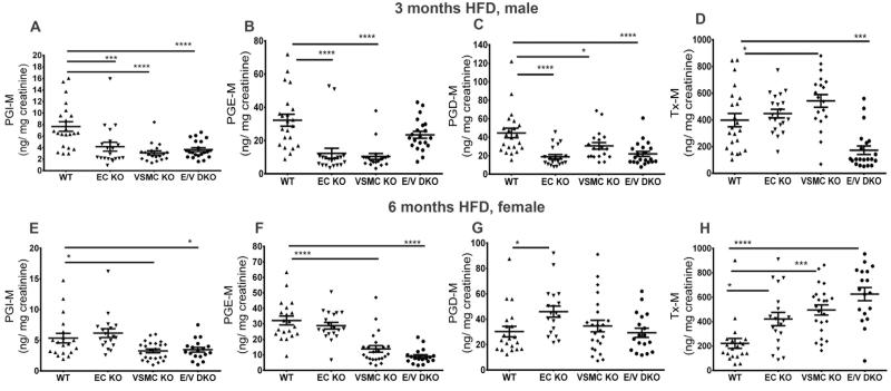
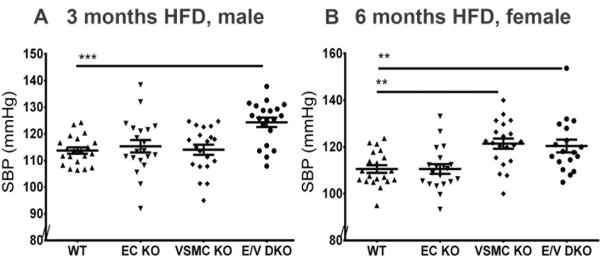
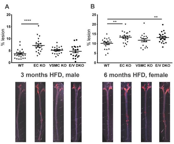
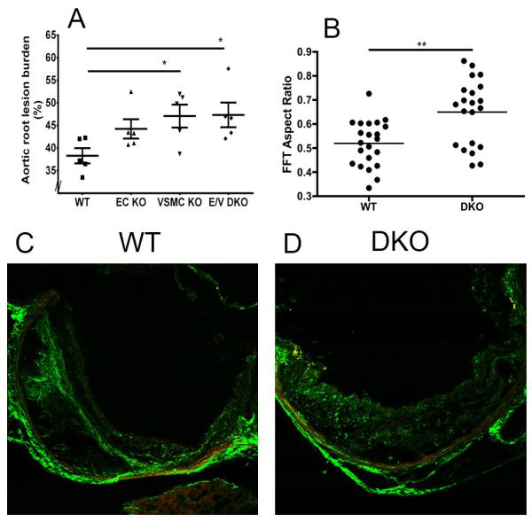
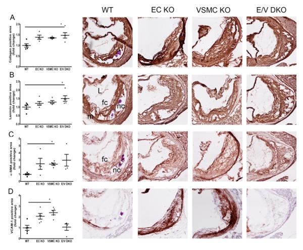
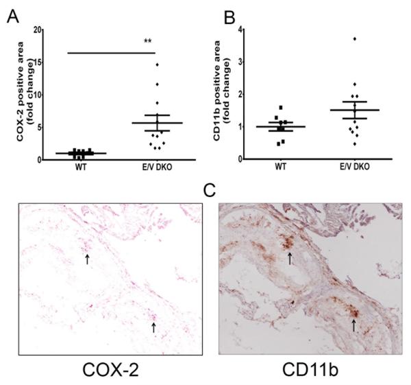
Methods and results: In the present study, selective depletion of COX-2 in vascular smooth muscle cells and endothelial cells depressed biosynthesis of prostaglandin I2 and prostaglandin E2, elevated blood pressure, and accelerated atherogenesis in Ldlr knockout mice. Deletion of COX-2 in vascular smooth muscle cells and endothelial cells coincided with an increase in COX-2 expression in lesional macrophages and increased biosynthesis of thromboxane. Increased accumulation of less organized intimal collagen, laminin, α-smooth muscle actin, and matrix-rich fibrosis was also apparent in lesions of the mutants.

Conclusions: Although atherogenesis is accelerated in global COX-2 knockouts, consistent with evidence of risk transformation during chronic nonsteroidal anti-inflammatory drug administration, this masks the contrasting effects of enzyme depletion in macrophages versus vascular smooth muscle cells and endothelial cells. Targeting delivery of COX-2 inhibitors to macrophages may conserve their efficacy while limiting cardiovascular risk.

Keywords: atherogenesis; cyclooxygenase; endothelial cell; prostaglandin; vascular smooth muscle.


Figures

Figure 1
Figure 1
Impact of COX-2 deletion on prostaglandin biosynthesis in mice on high fat diet. Overnight (5pm to 10am) fasting urine from WT and vascular COX-2 mutants were collected at the end of HFD feeding, and prostanoid metabolites were analyzed by liquid chromatography-mass spectrometry as described in the Methods. Urinary PGI-M (A and E) and PGE-M (B and F) were depressed in male and female COX-2 KOs after 3 and 6 months on HFD, respectively. Tx-M was increased in female KOs fed 6 months HFD. One-way ANOVA showed a significant effect of genotype on urinary prostanoid levels in both genders fed a HFD. Holm Sidak’s multiple comparison tests were used to test significant differences between WT and COX-2 mutants. Data are means ± SEMs. *p< 0.05, ***p<0.001, ****p<0.0001; n=18-22 per genotype.
Figure 2
Figure 2
Vascular specific COX-2 deletion elevates systolic blood pressure. Systolic blood pressure (SBP) was measured in conscious mice using the computerized non-invasive tail-cuff system as described in the Methods. SBP of male mice was elevated after 3 months on a HFD in E/V DKO compared to WT mice (A). This hypertensive phenotype was further augmented in all mutants after 6 months on a HFD, with the exception of female EC KOs (B). One-way ANOVA showed a significant effect of genotype on SBP in both genders. Holm Sidak’s multiple comparison tests were used to test significant differences between WT and COX-2 mutants. Data are means ± SEMs. **p< 0.01, ***p< 0.001; n=18-22 per genotype.
Figure 3
Figure 3
Vascular COX-2 restrains atherogenesis. Aortic atherosclerotic lesion burden, represented by the percentage of lesion area to total aortic area, was quantified by en face analysis of aortas from male mice fed a HFD for 3 and of female mice at 6 months. Representative en face preparations are shown (Lower panels). Lesion area tended to increase in male or female COX-2 mutants fed a HFD for 3 or 6 months respectively (A and B). One-way ANOVA revealed a significant effect of genotype (male, P= 0.0001 and female, P= 0.003) on lesion progression. Holm Sidak’s multiple comparison tests were used to test significant differences between WT and COX-2 KOs. Data are means ± SEMs. **p< 0.01, ****p<0.0001; n=18-22 per genotype.
Figure 4
Figure 4
Vascular COX-2 restrains aortic root lesion burden and orientation of structural collagen in lesions. A. Quantification of cross-sectional analysis of aortic root samples from female mice fed 6 months HFD, was performed by measuring total lesion area across the aortic root as detailed in Methods. One-way ANOVA (Kruskal-Wallis test) revealed a significant effect of genotype (P=0.048) on lesion progression. Dunnett’s multiple comparison tests were used to test significant differences between WT and COX-2 KOs. *p< 0.05. Data are means ± SEMs. n=5 per group and 7-9 sections from each animal were averaged. B. Second harmonic generation (SHG) detection of structural collagen in WT (n=22) and E/V DKO (n= 21) lesions from female mice on a HFD for 6 months. Fast Fourier transform (FFT)-based analyses revealed a more random orientation of collagen fibers in E/V DKO lesions. Data are means ± SEMs. **p < 0.01 (Mann-Whitney test, two-tails). C and D. Representative WT and E/V DKO SHG images are shown.
Figure 5
Figure 5
Morphometric consequences of vascular COX-2 deletion on lesion development. Lesion morphology in aortic roots from female mice fed HFD for 6 months were analyzed. Quantification of immunohistochemical staining of collagen (A), laminin (B), α-SMA (C) and VCAM-1 (D) from each genotype are shown in parallel with their representative aortic root sections. One-way ANOVA (Kruskal-Wallis test) revealed a significant effect of genotype (P<0.05) on lesion morphology. Dunnett’s multiple comparison tests were used to test significant differences between WT and COX-2 KOs. Data are means ± SEMs. n=4-5 per genotype. L- lumen, m- media, nc- necrotic core, fc- foam cell.
Figure 6
Figure 6
Vascular COX-2 deletion increases COX-2 expression in lesional macrophages. Lesion morphology in aortic roots from female mice fed a HFD for 6 months were analyzed. Quantification of immunohistochemistry staining of COX-2 (A) and CD11b (B) from WT and E/V DKO are shown. A Mann-Whitney test (two-tailed) revealed a significant increase in COX-2 positive cells in DKO compared to WT, **p< 0.01; n=8-12 per genotype. Data are means ± SEMs. C. Representative aortic root sections from COX-2 staining (left panel) and CD11b (right panel) from E/V DKO are shown. Arrows indicate lesional cells that are stained positive for both COX-2 and CD11b. L- lumen, m- media.

References

    1. FitzGerald GA. Cox-2 in play at the aha and the fda. Trends Pharmacol Sci. 2007;28:303–307. - PubMed
    1. Ricciotti E, FitzGerald GA. Prostaglandins and inflammation. Arterioscler Thromb Vasc Biol. 2011;31:986–1000. - PMC - PubMed
    1. Bertagnolli MM, Eagle CJ, Zauber AG, Redston M, Solomon SD, Kim K, Tang J, Rosenstein RB, Wittes J, Corle D, Hess TM, Woloj GM, Boisserie F, Anderson WF, Viner JL, Bagheri D, Burn J, Chung DC, Dewar T, Foley TR, Hoffman N, Macrae F, Pruitt RE, Saltzman JR, Salzberg B, Sylwestrowicz T, Gordon GB, Hawk ET, Investigators APCS Celecoxib for the prevention of sporadic colorectal adenomas. N Engl J Med. 2006;355:873–884. - PubMed
    1. Bresalier RS, Sandler RS, Quan H, Bolognese JA, Oxenius B, Horgan K, Lines C, Riddell R, Morton D, Lanas A, Konstam MA, Baron JA, Adenomatous Polyp Prevention on Vioxx Trial I Cardiovascular events associated with rofecoxib in a colorectal adenoma chemoprevention trial. N Engl J Med. 2005;352:1092–1102. - PubMed
    1. Solomon SD, McMurray JJ, Pfeffer MA, Wittes J, Fowler R, Finn P, Anderson WF, Zauber A, Hawk E, Bertagnolli M, Adenoma Prevention with Celecoxib Study I Cardiovascular risk associated with celecoxib in a clinical trial for colorectal adenoma prevention. N Engl J Med. 2005;352:1071–1080. - PubMed

Publication types

MeSH terms