

Ndfip1 mediates peripheral tolerance to self and exogenous antigen by inducing cell cycle exit in responding CD4+ T cells
- PMID: 24520172
- PMCID: PMC3926078
- DOI: 10.1073/pnas.1322739111
Ndfip1 mediates peripheral tolerance to self and exogenous antigen by inducing cell cycle exit in responding CD4+ T cells
Abstract
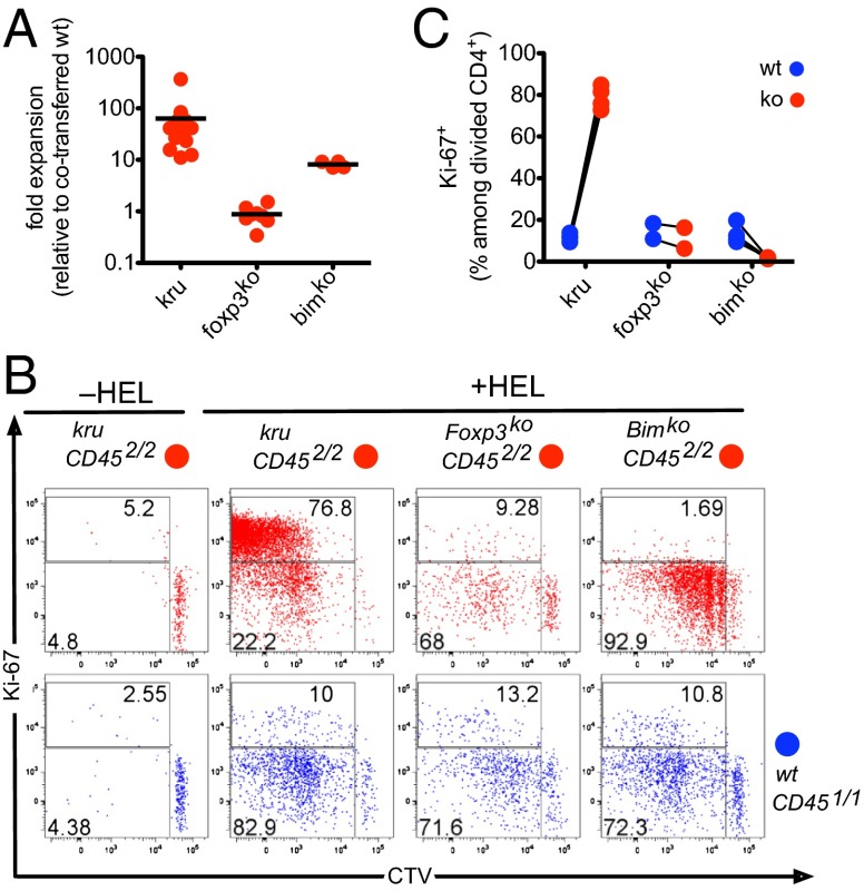
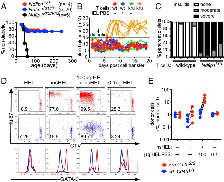
The NDFIP1 (neural precursor cell expressed, developmentally down-regulated protein 4 family-interacting protein 1) adapter for the ubiquitin ligase ITCH is genetically linked to human allergic and autoimmune disease, but the cellular mechanism by which these proteins enable foreign and self-antigens to be tolerated is unresolved. Here, we use two unique mouse strains--an Ndfip1-YFP reporter and an Ndfip1-deficient strain--to show that Ndfip1 is progressively induced during T-cell differentiation and activation in vivo and that its deficiency causes a cell-autonomous, Forkhead box P3-independent failure of peripheral CD4(+) T-cell tolerance to self and exogenous antigen. In small cohorts of antigen-specific CD4(+) cells responding in vivo, Ndfip1 was necessary for tolerogen-reactive T cells to exit cell cycle after one to five divisions and to abort Th2 effector differentiation, defining a step in peripheral tolerance that provides insights into the phenomenon of T-cell anergy in vivo and is distinct from the better understood process of Bcl2-interacting mediator of cell death-mediated apoptosis. Ndfip1 deficiency precipitated autoimmune pancreatic destruction and diabetes; however, this depended on a further accumulation of nontolerant anti-self T cells from strong stimulation by exogenous tolerogen. These findings illuminate a peripheral tolerance checkpoint that aborts T-cell clonal expansion against allergens and autoantigens and demonstrate how hypersensitive responses to environmental antigens may trigger autoimmunity.
Keywords: Aire (Autoimmune Regulator); Interleukin-4; T lymphocyte; allergy; immunological tolerance.
Conflict of interest statement
The authors declare no conflict of interest.
Figures
Similar articles
-
The Ubiquitin Ligase Adaptor NDFIP1 Selectively Enforces a CD8+ T Cell Tolerance Checkpoint to High-Dose Antigen.Cell Rep. 2018 Jul 17;24(3):577-584. doi: 10.1016/j.celrep.2018.06.060. Cell Rep. 2018. PMID: 30021156 Free PMC article.
-
Ndfip1 enforces a requirement for CD28 costimulation by limiting IL-2 production.J Immunol. 2013 Aug 15;191(4):1536-46. doi: 10.4049/jimmunol.1203571. Epub 2013 Jul 12. J Immunol. 2013. PMID: 23851689 Free PMC article.
-
TGF-β induces the expression of the adaptor Ndfip1 to silence IL-4 production during iTreg cell differentiation.Nat Immunol. 2011 Nov 13;13(1):77-85. doi: 10.1038/ni.2154. Nat Immunol. 2011. PMID: 22080920 Free PMC article.
-
Thymic commitment of regulatory T cells is a pathway of TCR-dependent selection that isolates repertoires undergoing positive or negative selection.Curr Top Microbiol Immunol. 2005;293:43-71. doi: 10.1007/3-540-27702-1_3. Curr Top Microbiol Immunol. 2005. PMID: 15981475 Review.
-
Research progress on the role of Ndfip1 (Nedd4 family interacting protein 1) in immune cells.Allergol Immunopathol (Madr). 2023 Jan 1;51(1):77-83. doi: 10.15586/aei.v51i1.739. eCollection 2023. Allergol Immunopathol (Madr). 2023. PMID: 36617825 Review.
Cited by
-
The Nedd4-2/Ndfip1 axis is a negative regulator of IgE-mediated mast cell activation.Nat Commun. 2016 Oct 27;7:13198. doi: 10.1038/ncomms13198. Nat Commun. 2016. PMID: 27786273 Free PMC article.
-
Functional characterization of eQTLs and asthma risk loci with scATAC-seq across immune cell types and contexts.Am J Hum Genet. 2025 Feb 6;112(2):301-317. doi: 10.1016/j.ajhg.2024.12.017. Epub 2025 Jan 14. Am J Hum Genet. 2025. PMID: 39814021 Free PMC article.
-
Identifying disease-critical cell types and cellular processes by integrating single-cell RNA-sequencing and human genetics.Nat Genet. 2022 Oct;54(10):1479-1492. doi: 10.1038/s41588-022-01187-9. Epub 2022 Sep 29. Nat Genet. 2022. PMID: 36175791 Free PMC article.
-
The Ubiquitin Ligase Adaptor NDFIP1 Selectively Enforces a CD8+ T Cell Tolerance Checkpoint to High-Dose Antigen.Cell Rep. 2018 Jul 17;24(3):577-584. doi: 10.1016/j.celrep.2018.06.060. Cell Rep. 2018. PMID: 30021156 Free PMC article.
-
The CD8+ T cell tolerance checkpoint triggers a distinct differentiation state defined by protein translation defects.Immunity. 2024 Jun 11;57(6):1324-1344.e8. doi: 10.1016/j.immuni.2024.04.026. Epub 2024 May 21. Immunity. 2024. PMID: 38776918 Free PMC article.
References
-
- Goodnow CC, Ohashi PS. 2013. Immunologic Tolerance. Fundamental Immunology, ed Paul WE, 7th Ed (Wolters Kluwer Health/Lippincott Williams and Wilkins, Philadelphia), pp 765–794.
-
- Harvey KF, Shearwin-Whyatt LM, Fotia A, Parton RG, Kumar S. N4WBP5, a potential target for ubiquitination by the Nedd4 family of proteins, is a novel Golgi-associated protein. J Biol Chem. 2002;277(11):9307–9317. - PubMed
Publication types
MeSH terms
Substances
Grants and funding
LinkOut - more resources
Full Text Sources
Other Literature Sources
Molecular Biology Databases
Research Materials
