. 2014 Mar 4;111(9):3544-9.
doi: 10.1073/pnas.1314118111. Epub 2014 Feb 11.

Tissue factor expression provokes escape from tumor dormancy and leads to genomic alterations

Affiliations

Tissue factor expression provokes escape from tumor dormancy and leads to genomic alterations

Nathalie Magnus et al. Proc Natl Acad Sci U S A. .

Abstract

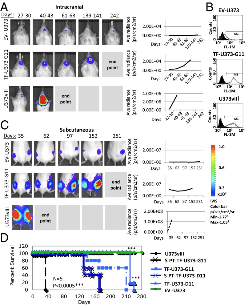
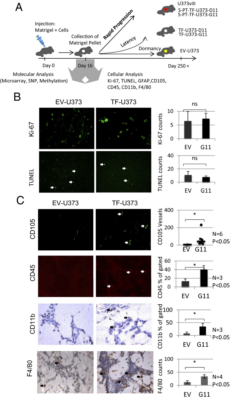
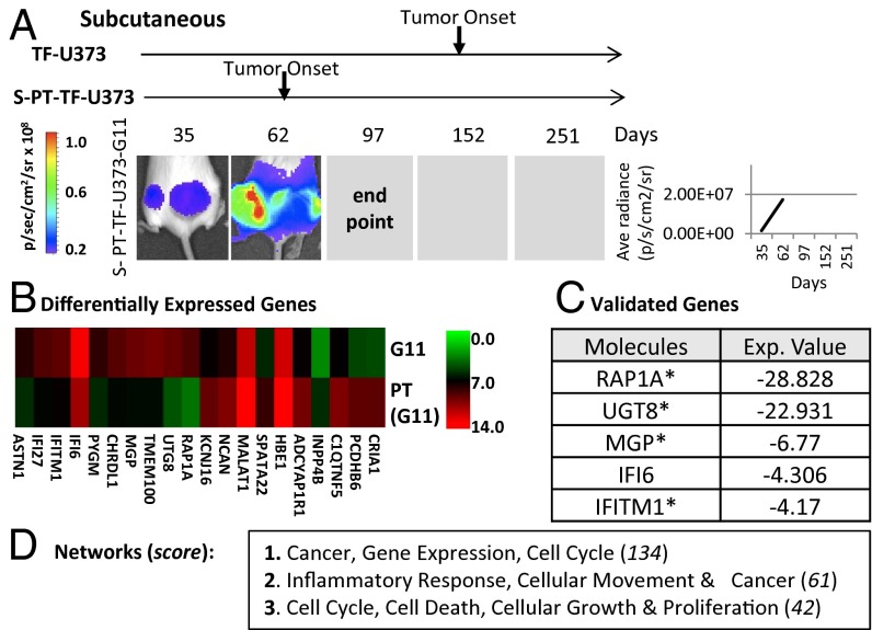
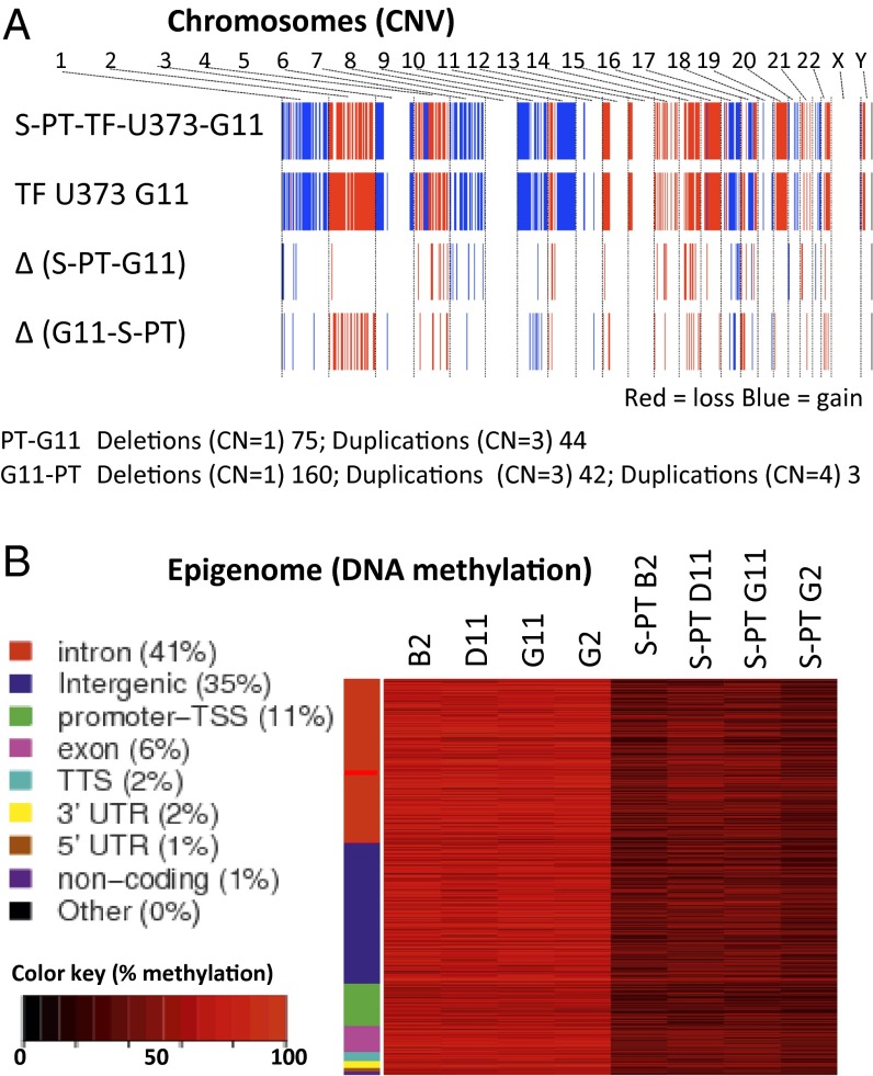
The coagulation system links immediate (hemostatic) and late (inflammatory, angiogenic) tissue responses to injury, a continuum that often is subverted in cancer. Here we provide evidence that tumor dormancy is influenced by tissue factor (TF), the cancer cell-associated initiator of the coagulation system and a signaling receptor. Thus, indolent human glioma cells deficient for TF remain viable but permanently dormant at the injection site for nearly a year, whereas the expression of TF leads to a step-wise transition to latent and overt tumor growth phases, a process that is preceded by recruitment of vascular (CD105(+)) and myeloid (CD11b(+) and F4/80(+)) cells. Importantly, the microenvironment orchestrated by TF expression drives permanent changes in the phenotype, gene-expression profile, DNA copy number, and DNA methylation state of the tumor cells that escape from dormancy. We postulate that procoagulant events in the tissue microenvironment (niche) may affect the fate of occult tumor cells, including their biological and genetic progression to initiate a full-blown malignancy.

Keywords: angiogenesis; brain tumor; clotting; macrophages; oncogenes.


Conflict of interest statement

The authors declare no conflict of interest.

Figures

Fig. 1.
Fig. 1.
TF expression drives the escape of tumors from dormancy. (A) Dormant phenotype of EV-U373 cells is interrupted by the expression of TF. (Left) Bioluminescent imaging of Luciferase-expressing cell lines upon orthotopic injection into SCID mice. TF-U373-G11 cells injected intracranially remain latent but eventually emerge as detectable tumors. (Right) Quantitative plots of all tumors included in these experiments. (B) Flow cytometry shows immunostaining for the cell-surface expression of TF in parental/control cells (EV-U373), their derivatives expressing EGFRvIII (U373vIII), and an example of the TF transfectant clone (TF-U373-G11); compare with data in SI Appendix, Table S1. (C) Dormant phenotype of EV-U373 cells inoculated s.c. is interrupted by the enforced expression of TF. (Left) Bioluminescent images of s.c. tumors that were generated by EV-373, TF-U373-G11, and U373vIII cells. (Right) Cumulative growth plots combining all mice in the respective groups. (D) Impact of TF expression on survival of mice harboring dormant and aggressive glioma xenografts. Representative Kaplan–Meier plots depict survival of mice; n = 5 per group. EV-U373 cells remain dormant; TF-U373-G11 and TF-U373-D11 cell lines engineered to express TF form tumors after a long latency and with less than 100% tumor take rate (SI Appendix). PT, primary tumor cells derived by culturing cells isolated from TF-U373 tumors. Significance of differences was assessed by log-rank test; ***P < 0.0005.
Fig. 2.
Fig. 2.
Angiogenesis and inflammation precede TF-driven tumor escape from dormancy. (A) Diagram showing the analysis of molecular and cellular changes preceding the escape from tumor dormancy. Tumor cells embedded in Matrigel were injected on day 0, and the pellets were retrieved on day 16. The cellular content of pellets was analyzed for cellular and molecular parameters as indicated (see text). (B) Staining of tumor inoculates for markers of proliferation (KI67) and apoptosis (TUNEL). EV-U373 and TF-U373-G11 pellets contain comparable numbers of dividing and apoptotic cells. (Upper) Quantification of KI67 at 40× magnification; n = 4; P > 0.05. (Lower) TUNEL staining; n = 4; P > 0.05. (C) Changes in the content of endothelial and leukocytic cells as a function of TF expression by glioma cells. (Top Row) Immunohistochemistry for CD105 reveals a differential influx of endothelial cells and blood vessels into Matrigel pellets containing EV-U373 and TF-U373-G11 cells. (Right) Quantification of CD105-positive vascular structures, n = 6; *P < 0.05. (Second Row) (Left) Immunohistochemistry for CD45+ cells (arrows). (Right) Flow cytometry quantification of CD45+ leukocytic cells within Matrigel pellets. n = 3; *P < 0.05. (Third Row) (Left) Immunostaining for CD11b+ myeloid cells in tumor cell-containing Matrigel pellets (black arrows). (Right) Flow cytometry quantification of CD11b+ cells in Matrigel pellets. n = 3; *P < 0.05. (Bottom Row) (Left) Immunostaining for the F4/80 marker of macrophages. (Right) Quantification of the F4/80 signal in pellets n = 4; *P < 0.05.
Fig. 3.
Fig. 3.
Growth in the TF-controlled microenvironment provokes permanent changes in cellular phenotype. (A) Expression of a highly tumorigenic phenotype in TF-expressing glioma cells after their passage as tumors in SCID mice. (Left) Bioluminescent images of s.c. primary tumors (S-PT-TF-U373-G11); arrows indicate the time of tumor onset in various experiments (SI Appendix). (Right) Line graph summarizing of tumor growth for all mice in the group (n = 5). (B) Changes in the gene-expression profile between TF-U373-G11 cells and their tumor-derived variant (S-PT-U373-G11). The heatmap generated from the output of the human Affymetrix U133 array compares the levels of 10 most different mRNA species between TF-U373-G11 and S-PT-TF-U373-G11 cells embedded in Matrigel pellets in vivo (day 16 postinjection). (C) List of genes validated by semiquantitative RT-PCR. (D) Table showing the top three networks according to the IPA software, including genes with a greater than twofold change.
Fig. 4.
Fig. 4.
Growth of glioma cells in the TF-controlled microenvironment drives permanent changes in cellular genome and epigenome. (A) CNV differences between TF-U373-G11 cells and their derived isogenic variant S-PT-U373-G11 generated in vivo (see text). SNP analysis (Affymetrix 6.0) documents altered regions of genomic DNA. (B) Cellular methylome profiling using the RRBS strategy between indicated cell lines cultured in vitro. Hyper- and hypomethylated tiles with a greater than 20% difference in methylation between conditions and Fisher’s exact test (q-value <0.01) were used to create the heatmap. There is a clear separation between the GpC methylation profiles of several independent TF-expressing clonal cell lines and their tumor-derived counterparts (the PT series of cell lines).

References

    1. Folkman J, Kalluri R. Cancer without disease. Nature. 2004;427(6977):787. - PubMed
    1. Aguirre-Ghiso JA. Models, mechanisms and clinical evidence for cancer dormancy. Nat Rev Cancer. 2007;7(11):834–846. - PMC - PubMed
    1. Almog N. Molecular mechanisms underlying tumor dormancy. Cancer Lett. 2010;294(2):139–146. - PubMed
    1. Black WC, Welch HG. Advances in diagnostic imaging and overestimations of disease prevalence and the benefits of therapy. N Engl J Med. 1993;328(17):1237–1243. - PubMed
    1. Goss PE, Chambers AF. Does tumour dormancy offer a therapeutic target? Nat Rev Cancer. 2010;10(12):871–877. - PubMed

Publication types