. 2014 Feb 10;9(2):e88511.
doi: 10.1371/journal.pone.0088511. eCollection 2014.

Novel brain arteriovenous malformation mouse models for type 1 hereditary hemorrhagic telangiectasia

Affiliations

Novel brain arteriovenous malformation mouse models for type 1 hereditary hemorrhagic telangiectasia

Eun-Jung Choi et al. PLoS One. .

Abstract

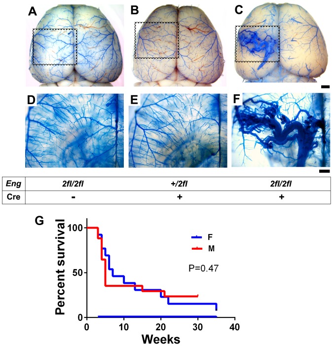
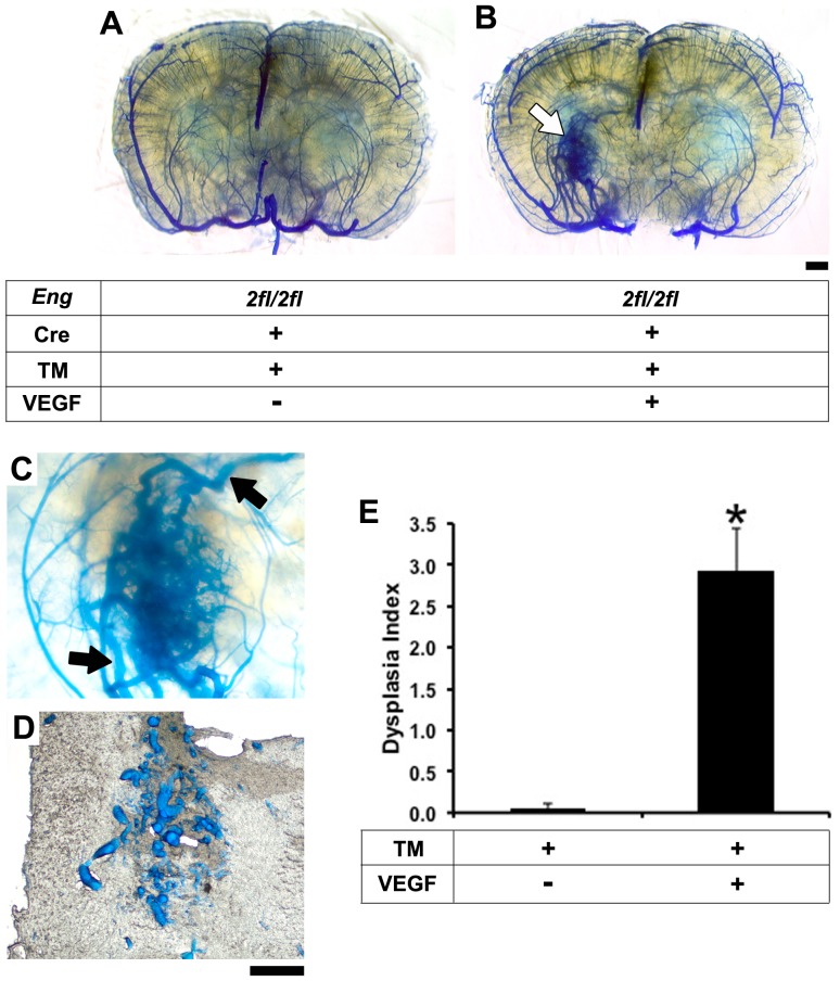
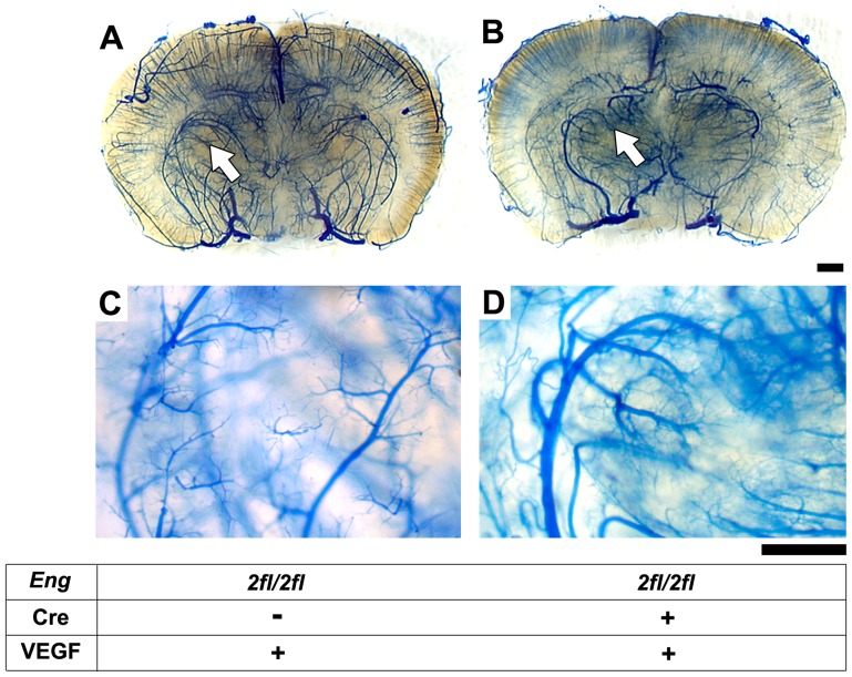
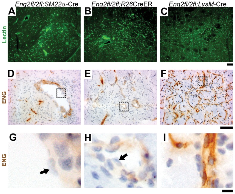
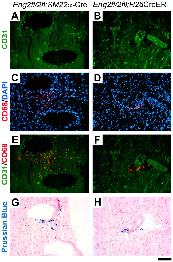
Endoglin (ENG) is a causative gene of type 1 hereditary hemorrhagic telangiectasia (HHT1). HHT1 patients have a higher prevalence of brain arteriovenous malformation (AVM) than the general population and patients with other HHT subtypes. The pathogenesis of brain AVM in HHT1 patients is currently unknown and no specific medical therapy is available to treat patients. Proper animal models are crucial for identifying the underlying mechanisms for brain AVM development and for testing new therapies. However, creating HHT1 brain AVM models has been quite challenging because of difficulties related to deleting Eng-floxed sequence in Eng(2fl/2fl) mice. To create an HHT1 brain AVM mouse model, we used several Cre transgenic mouse lines to delete Eng in different cell-types in Eng(2fl/2fl) mice: R26CreER (all cell types after tamoxifen treatment), SM22α-Cre (smooth muscle and endothelial cell) and LysM-Cre (lysozyme M-positive macrophage). An adeno-associated viral vector expressing vascular endothelial growth factor (AAV-VEGF) was injected into the brain to induce focal angiogenesis. We found that SM22α-Cre-mediated Eng deletion in the embryo caused AVMs in the postnatal brain, spinal cord, and intestines. Induction of Eng deletion in adult mice using R26CreER plus local VEGF stimulation induced the brain AVM phenotype. In both models, Eng-null endothelial cells were detected in the brain AVM lesions, and formed mosaicism with wildtype endothelial cells. However, LysM-Cre-mediated Eng deletion in the embryo did not cause AVM in the postnatal brain even after VEGF stimulation. In this study, we report two novel HHT1 brain AVM models that mimic many phenotypes of human brain AVM and can thus be used for studying brain AVM pathogenesis and testing new therapies. Further, our data indicate that macrophage Eng deletion is insufficient and that endothelial Eng homozygous deletion is required for HHT1 brain AVM development.


Conflict of interest statement

Competing Interests: The authors have declared that no competing interests exist.

Figures

Figure 1
Figure 1. Developmental onset AVM in the postnatal brain of Eng2fl/2fl;SM22α-Cre mice.
Representative images of latex dye casting show the cerebrovasculature in the brain of 5-week-old (A) Eng2fl/2fl, (B) Eng+/2fl;SM22α-Cre, and (C) Eng2fl/2fl;SM22α-Cre mice. D–F: Enlarged images of dotted boxes shown in A–C after the brain tissue was cleared in organic solvent. The latex-perfused vasculature inside the brain can be observed. Tangled and dilated vessels were detected only in the brain of Eng2fl/2fl;SM22α-Cre mice. Scale bars: 1 mm in A–C and 500 µm in D–F. G: Survival curve of Eng2fl/2fl;SM22α-Cre mice. There was no difference between male (M) and female (F) mice (P = 0.47).
Figure 2
Figure 2. Adult onset brain AVM in Eng2fl/2fl;R26CreER mice after TM and VEGF treatment.
A: Coronal view of latex-perfused adult Eng2fl/2fl;R26CreER brain showing no AVM phenotype 8 weeks after TM treatment. B: AVM phenotype (white arrow) developed in the brain of Eng2fl/2fl;R26CreER mice 8 weeks after intrabrain injection of AAV-VEGF and intraperitoneal injection of TM. C: An enlarged image of the AVM lesion. Latex-perfused veins are clearly shown at the top and bottom of the lesion (black arrows). D: Enlarged vessels observed in a 50 µm frozen section that cuts through the AVM lesion shown in C. E: Quantification of dysplasia index. Data: mean ± SD. *: p<0.05. n = 6 per group. Scale bars: 1 mm in A and B and 500 µm in C and D.
Figure 3
Figure 3. AVM phenotype was not detected in the brain angiogenic region of Eng2fl/2fl;LysM-Cre mice.
Representative images of the latex-casted cerebrovasculature in (A) Eng2fl/2fl and (B) Eng2fl/2fl;LysMCre adult mice 8 weeks after focal VEGF stimulation. C and D: High magnification view of the angiogenic foci (arrows) shown in A and B. Latex was present in arteries only. No vein and enlarged and tangled vessels were detected. Scale bars: 1 mm in A and B and 500 µm in C and D.
Figure 4
Figure 4. ENG-null endothelial cells in dysplastic vessels.
Representative images of lectin-stained brain sections from (A) AVM lesion of 5-week-old Eng2fl/2fl;SM22α-Cre, (B) VEGF-induced angiogenic focus of TM-treated adult Eng2fl/2fl;R26CreER, and (C) VEGF-stimulated angiogenic focus of adult Eng2fl/2fl;LysM-Cre mice. ENG expression in (D) Eng2fl/2fl;SM22α-Cre, (E) Eng2fl/2fl;R26CreER, and (F) Eng2fl/2fl;LysM-Cre brain. G-I: Enlarged images of the dotted boxes shown in D-F. Arrows indicate ENG-negative endothelial cells. Scale bars: 100 µm in A–F and 10 µm in G–I.
Figure 5
Figure 5. Macrophage infiltration and microhemorrhage in brain AVM lesions of Eng2fl/2fl;SM22αCre and Eng2fl/2fl;R26CreER mice.
Dysplastic vessels were present in the brain of (A) Eng2fl/2fl;SM22α-Cre and (B) Eng2fl/2fl;R26CreER mice. Endothelial cells were visualized by immunostaining using an antibody specific to CD31 (an endothelial cell-specific marker). C and D: Macrophages were detected by immunostaining using a CD68-specific antibody. The nuclei were stained by DAPI. E and F: Macrophages localized around large dysplastic vessels. G and H: Iron deposits, an indicator of hemorrhage, identified by Prussian blue. Scale bar: 100 µm.

References

    1. McAllister KA, Grogg KM, Johnson DW, Gallione CJ, Baldwin MA, et al. (1994) Endoglin, a TGF-beta binding protein of endothelial cells, is the gene for hereditary haemorrhagic telangiectasia type 1. Nat Genet 8: 345–351. - PubMed
    1. Shovlin CL, Guttmacher AE, Buscarini E, Faughnan ME, Hyland RH, et al. (2000) Diagnostic criteria for hereditary hemorrhagic telangiectasia (Rendu-Osler-Weber syndrome). Am J Med Genet 91: 66–67. - PubMed
    1. Johnson DW, Berg JN, Baldwin MA, Gallione CJ, Marondel I, et al. (1996) Mutations in the activin receptor-like kinase 1 gene in hereditary haemorrhagic telangiectasia type 2. Nat Genet 13: 189–195. - PubMed
    1. Kim H, Marchuk DA, Pawlikowska L, Chen Y, Su H, et al... (2008) Genetic considerations relevant to intracranial hemorrhage and brain arteriovenous malformations. Acta Neurochir Suppl 105: 199–206. - PMC - PubMed
    1. Kim H, Su H, Weinsheimer S, Pawlikowska L, Young WL (2011) Brain arteriovenous malformation pathogenesis: a response-to-injury paradigm. Acta Neurochir Suppl 111: 83–92. - PMC - PubMed

Publication types

MeSH terms