

Reproductive performance of mouse oocyte after in vivo exposure of the ovary to continuous wave ultrasound
- PMID: 24520439
- PMCID: PMC3850303
Reproductive performance of mouse oocyte after in vivo exposure of the ovary to continuous wave ultrasound
Abstract
Background: There is a lack of studies regarding the effects of ultrasound (US) and replication of its exposure on pre-implantation events in mammals. Thus, this study assesses the reproductive performance of mouse oocytes that have been obtained from ovaries irradiated with US waves versus non-irradiated ovaries. Also comparision of their parthenogenesis, ovulation, fertilization, and pre-implantation development rates.
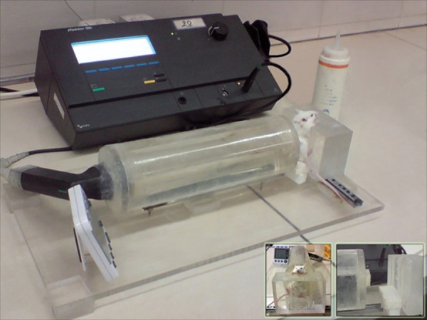
Materials and methods: In this experimental study, we divided extracted ovaries into three experimental groups that received the same dosage, but different replicates of radiation for each group. Results were compared with the control and sham groups. Continuous wave (CW) US, at a spatial average intensity of 355 mW/cm(2) and a frequency of 3.28 MHz, was administered for 5 minutes to the ovaries at an interval between pregnant mare serum gonadotropin (PMSG) and human chorionic gonadotropin (hCG) injections. Statistical analysis was performed using the ANOVA test and the level of significance was determined to be 0.05.
Results: Data collection was based on microscopic visualization. According to the obtained results, metaphase II (MII) oocyte numbers and the percentage of blastocysts significantly reduced in the USexposed groups versus the unexposed groups. Fertilization rate was comparable between groups while parthenogenesis was significantly higher in the US-exposed groups compared to the unexposed groups.
Conclusion: Structural damage to cells, intracellular organelles and proteins, as well as changes in signaling pathways induced by US may be reasons for some of the observed adverse effects in groups that have received more US exposure.
Keywords: Blastocyst; Fertilization Rate; Mouse Oocyte; Parthenogenesis; Ultrasound.
Figures
References
-
- Baker KG, Robertson VJ, Duck FA. A review of therapeutic ultrasound: biophysical effects. Phys Ther. 2001;81(7):1351–1358. - PubMed
-
- Bailey MR, Khokhlova VA, Sapozhnikov OA, Kargl SG, Crum LA. Physical mechanisms of the therapeutic effect of ultrasound. Acoust Phys. 2003;49(4):369–388.
-
- Heyner S, Abraham V, Wicarszuk ML, ZiSkin MC. Effect of ultrasound on ovulation in the mouse. Gamete Res. 1989;22(3):333–338. - PubMed
-
- Testart J, Thebaoult A, Souderes E, Frydman R. Premature ovulation after ovarian ultrasonography. Br J Obstet Gynaecol. 1982;89(9):694–700. - PubMed
-
- Miyoshi K, Fujimoto Y, Mori H, Yoshida M. Activation and parthenogenetic development of pig oocytes exposed to ultrasound in media containing different concentration of ca2+ J Reprod Dev. 2008;54(1):42–45. - PubMed
LinkOut - more resources
Full Text Sources