

Balloon cell melanoma: a case report with polarized and non-polarized dermatoscopy and dermatopathology
- PMID: 24520518
- PMCID: PMC3919844
- DOI: 10.5826/dpc.0401a11
Balloon cell melanoma: a case report with polarized and non-polarized dermatoscopy and dermatopathology
Abstract
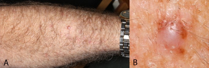
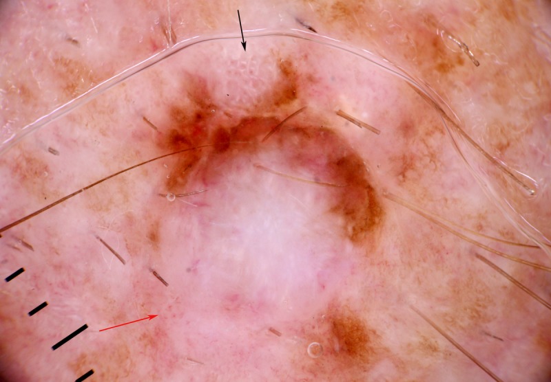
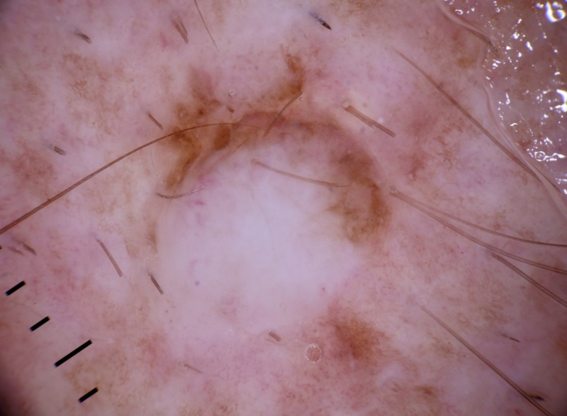
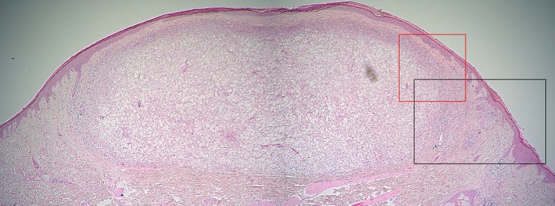
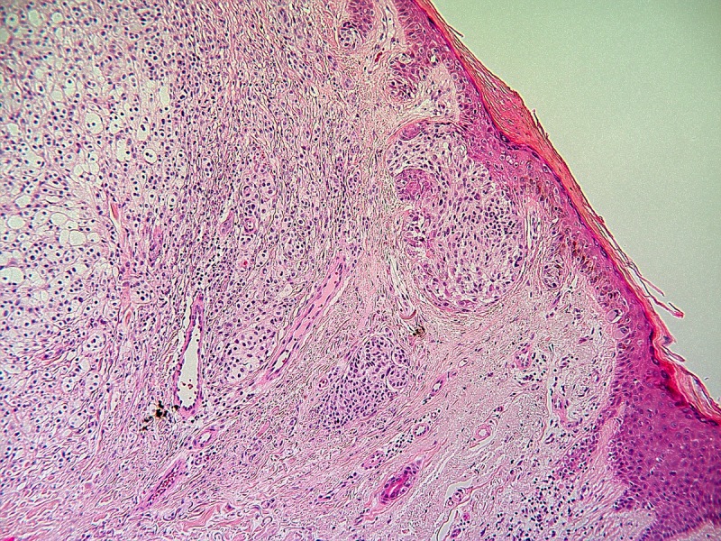
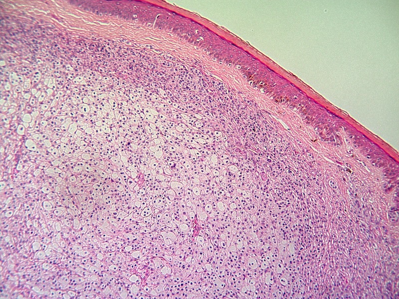
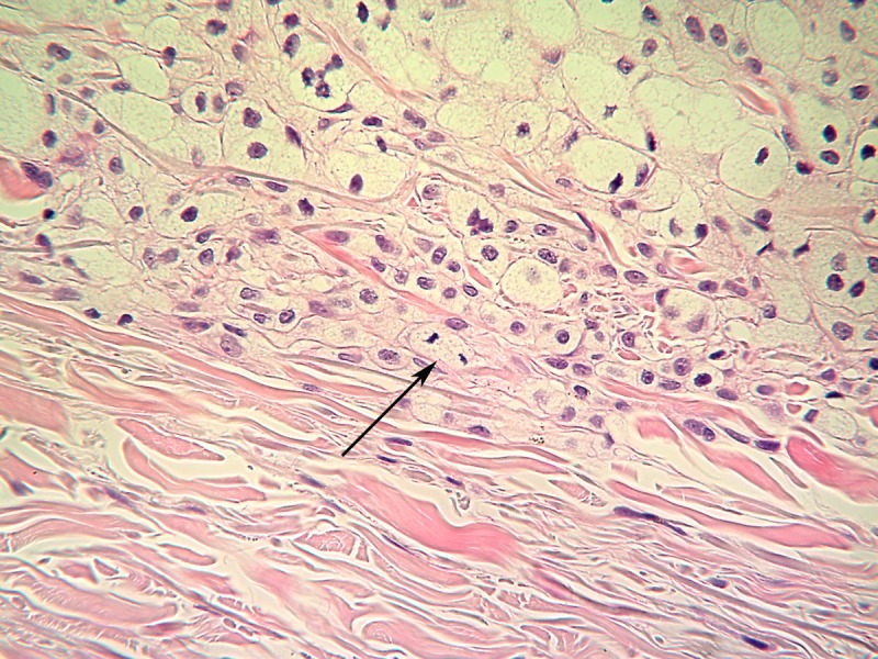
Balloon cell melanoma is a rare melanoma subtype, with only one previous case with dermatoscopy published. It is often non-pigmented, leading to diagnostic difficulty, and there is a tendency for lesions to be thick at diagnosis. We report a case of balloon cell melanoma on the forearm of a 61-year-old man with both polarized and non-polarized dermatoscopy and dermatopathology. It presented as a firm pale nodule with focal eccentric pigmentation. The clinical images evoke a differential diagnosis of dermatofibroma, dermal nevus, Spitz nevus and basal cell carcinoma as well as melanoma. This melanoma was partially pigmented due to a small, pigmented superficial spreading component on the edge of the non-pigmented balloon cell nodule, prompting further evaluation. In retrospect there was the clue to malignancy of polarizing-specific white lines (chrysalis structures) and polymorphous vessels, including a pattern of dot vessels. The reticular lines exclude basal cell carcinoma, polarizing-specific white lines are inconsistent with the diagnosis of dermal nevus and their eccentric location is inconsistent with both Spitz nevus and dermatofibroma. Excision biopsy was performed, revealing a superficial spreading melanoma with two distinct invasive components, one of atypical non-mature epithelioid cells and the other an amelanotic nodular component, comprising more than 50% of the lesion, characterized by markedly distended epithelioid melanocytes showing pseudo-xanthomatous cytoplasmic balloon cell morphology. A diagnosis of balloon cell melanoma, Breslow thickness 1.9 mm, mitotic rate 3 per square millimeter was rendered. Wide local excision was performed, as was sentinel lymph node biopsy, which was negative.
Keywords: balloon cell melanoma; balloon cells; chrysalis structures; dermatopathology; dermatoscopy; dermoscopy.
Figures
Similar articles
-
Glowing in the dark: case report of a clue-poor melanoma unmasked by polarized dermatoscopy.Dermatol Pract Concept. 2014 Jan 31;4(1):83-7. doi: 10.5826/dpc.0401a14. eCollection 2014 Jan. Dermatol Pract Concept. 2014. PMID: 24520521 Free PMC article.
-
Balloon cell melanoma in primary care practice: a case report.Dermatol Pract Concept. 2013 Jul 31;3(3):25-9. doi: 10.5826/dpc.0303a08.. eCollection 2013. Dermatol Pract Concept. 2013. PMID: 24106659 Free PMC article.
-
Nodular melanoma: five consecutive cases in a general practice with polarized and non-polarized dermatoscopy and dermatopathology.Dermatol Pract Concept. 2014 Apr 30;4(2):69-75. doi: 10.5826/dpc.0402a15. eCollection 2014 Apr. Dermatol Pract Concept. 2014. PMID: 24855580 Free PMC article.
-
Role of In Vivo Reflectance Confocal Microscopy in the Analysis of Melanocytic Lesions.Acta Dermatovenerol Croat. 2018 Apr;26(1):64-67. Acta Dermatovenerol Croat. 2018. PMID: 29782304 Review.
-
Clinical and Dermoscopic Features Associated With Difficult-to-Recognize Variants of Cutaneous Melanoma: A Systematic Review.JAMA Dermatol. 2020 Apr 1;156(4):430-439. doi: 10.1001/jamadermatol.2019.4912. JAMA Dermatol. 2020. PMID: 32101255
Cited by
-
Non-choroidal yellow melanoma showing positive staining with Sudan Black consistent with the presence of lipofuscin: a case report.Dermatol Pract Concept. 2014 Apr 30;4(2):45-9. doi: 10.5826/dpc.0402a09. eCollection 2014 Apr. Dermatol Pract Concept. 2014. PMID: 24855574 Free PMC article.
-
Association of Clinical, Dermoscopic, and Histopathologic Findings With Gene Expression in Patients With Balloon Cell Melanoma.JAMA Dermatol. 2018 Jan 1;154(1):77-81. doi: 10.1001/jamadermatol.2017.4700. JAMA Dermatol. 2018. PMID: 29238799 Free PMC article.
-
Balloon Cell Melanoma: Presentation of Four Cases with a Comprehensive Review of the Literature.Dermatopathology (Basel). 2022 Mar 28;9(2):100-110. doi: 10.3390/dermatopathology9020013. Dermatopathology (Basel). 2022. PMID: 35466242 Free PMC article. Review.
-
The diagnostic value and histologic correlate of distinct patterns of shiny white streaks for the diagnosis of melanoma: A retrospective, case-control study.J Am Acad Dermatol. 2018 May;78(5):913-919. doi: 10.1016/j.jaad.2017.11.021. Epub 2017 Nov 11. J Am Acad Dermatol. 2018. PMID: 29138058 Free PMC article.
-
The role of short-term, low dose intravenous ketamine infusion in Calciphylaxis.CEN Case Rep. 2021 Aug;10(3):422-425. doi: 10.1007/s13730-020-00557-8. Epub 2021 Feb 19. CEN Case Rep. 2021. PMID: 33606191 Free PMC article.
References
-
- Kao GF, Helwig EB, Graham JH. Balloon cell malignant melanoma of the skin. A clinicopathologic study of 34 cases with histochemical, immunohistochemical, and ultrastructural observations. Cancer. 1992;69(12):2942–52. 15. - PubMed
-
- Gardner WA, Jr, Vazquez MD. Balloon cell melanoma. Arch Pathol. 1970;89(5):470–2. - PubMed
-
- Magro CM, Crowson AN, Mihm MC. Unusual variants of malignant melanoma. Mod. Pathol. 2006;19(Suppl 2):S41–70. - PubMed
-
- Zaballos P, Puig S, Llambrich A, Malvehy J. Dermoscopy of dermatofibromas: a prospective morphological study of 412 cases. Arch Dermatol. 2008;144(1):75–83. - PubMed
Publication types
LinkOut - more resources
Full Text Sources
Other Literature Sources