

Balloon cell melanoma: a case report with polarized and non-polarized dermatoscopy and dermatopathology
- PMID: 24520518
- PMCID: PMC3919844
- DOI: 10.5826/dpc.0401a11
Balloon cell melanoma: a case report with polarized and non-polarized dermatoscopy and dermatopathology
Abstract
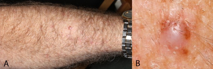
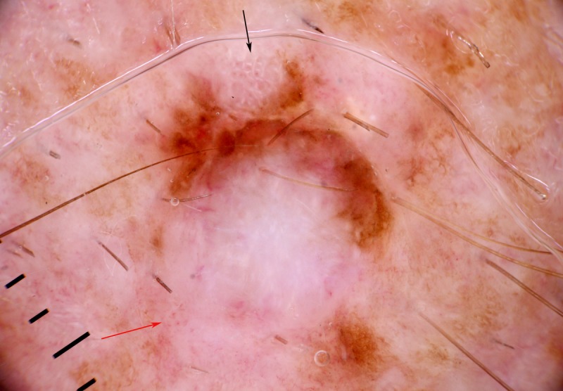
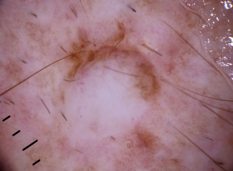
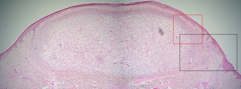
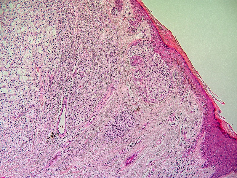
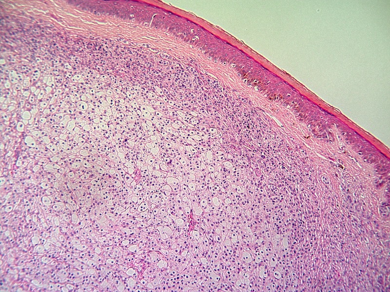
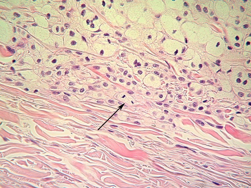
Balloon cell melanoma is a rare melanoma subtype, with only one previous case with dermatoscopy published. It is often non-pigmented, leading to diagnostic difficulty, and there is a tendency for lesions to be thick at diagnosis. We report a case of balloon cell melanoma on the forearm of a 61-year-old man with both polarized and non-polarized dermatoscopy and dermatopathology. It presented as a firm pale nodule with focal eccentric pigmentation. The clinical images evoke a differential diagnosis of dermatofibroma, dermal nevus, Spitz nevus and basal cell carcinoma as well as melanoma. This melanoma was partially pigmented due to a small, pigmented superficial spreading component on the edge of the non-pigmented balloon cell nodule, prompting further evaluation. In retrospect there was the clue to malignancy of polarizing-specific white lines (chrysalis structures) and polymorphous vessels, including a pattern of dot vessels. The reticular lines exclude basal cell carcinoma, polarizing-specific white lines are inconsistent with the diagnosis of dermal nevus and their eccentric location is inconsistent with both Spitz nevus and dermatofibroma. Excision biopsy was performed, revealing a superficial spreading melanoma with two distinct invasive components, one of atypical non-mature epithelioid cells and the other an amelanotic nodular component, comprising more than 50% of the lesion, characterized by markedly distended epithelioid melanocytes showing pseudo-xanthomatous cytoplasmic balloon cell morphology. A diagnosis of balloon cell melanoma, Breslow thickness 1.9 mm, mitotic rate 3 per square millimeter was rendered. Wide local excision was performed, as was sentinel lymph node biopsy, which was negative.
Keywords: balloon cell melanoma; balloon cells; chrysalis structures; dermatopathology; dermatoscopy; dermoscopy.
Figures
References
-
- Kao GF, Helwig EB, Graham JH. Balloon cell malignant melanoma of the skin. A clinicopathologic study of 34 cases with histochemical, immunohistochemical, and ultrastructural observations. Cancer. 1992;69(12):2942–52. 15. - PubMed
-
- Gardner WA, Jr, Vazquez MD. Balloon cell melanoma. Arch Pathol. 1970;89(5):470–2. - PubMed
-
- Magro CM, Crowson AN, Mihm MC. Unusual variants of malignant melanoma. Mod. Pathol. 2006;19(Suppl 2):S41–70. - PubMed
-
- Zaballos P, Puig S, Llambrich A, Malvehy J. Dermoscopy of dermatofibromas: a prospective morphological study of 412 cases. Arch Dermatol. 2008;144(1):75–83. - PubMed
Publication types
LinkOut - more resources
Full Text Sources
Other Literature Sources