. 2014 Mar 11;53(9):1536-43.
doi: 10.1021/bi401716z. Epub 2014 Feb 26.

Galactoside-binding site in LacY

Affiliations

Galactoside-binding site in LacY

Xiaoxu Jiang et al. Biochemistry. .

Abstract

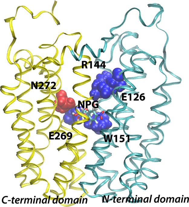
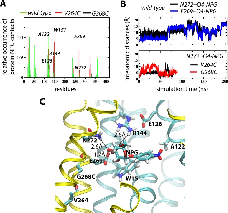
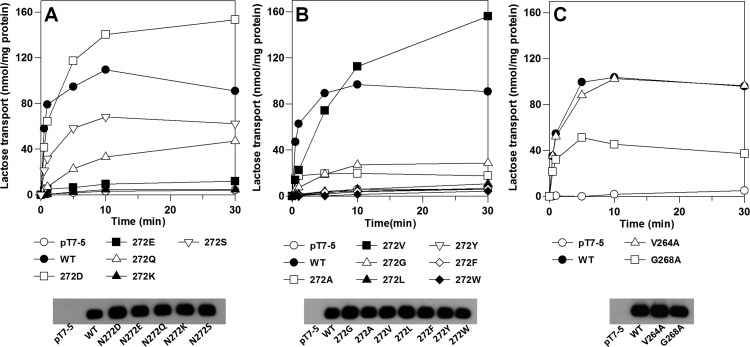
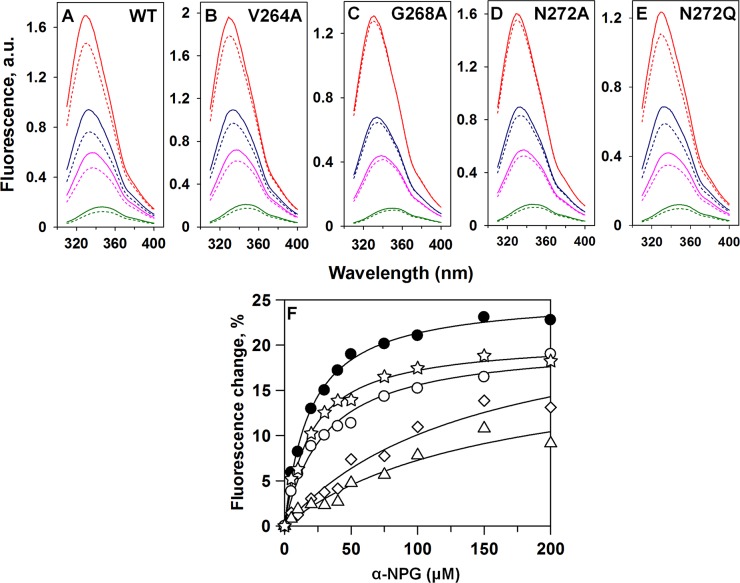
Although an X-ray crystal structure of lactose permease (LacY) has been presented with bound galactopyranoside, neither the sugar nor the residues ligating the sugar can be identified with precision at ~3.5 Å. Therefore, additional evidence is important for identifying side chains likely to be involved in binding. On the basis of a clue from site-directed alkylation suggesting that Asn272, Gly268, and Val264 on one face of helix VIII might participate in galactoside binding, molecular dynamics simulations were conducted initially. The simulations indicate that Asn272 (helix VIII) is sufficiently close to the galactopyranosyl ring of a docked lactose analogue to play an important role in binding, the backbone at Gly268 may be involved, and Val264 does not interact with the bound sugar. When the three side chains are subjected to site-directed mutagenesis, with the sole exception of mutant Asn272 → Gln, various other replacements for Asn272 either markedly decrease affinity for the substrate (i.e., high KD) or abolish binding altogether. However, mutant Gly268 → Ala exhibits a moderate 8-fold decrease in affinity, and binding by mutant Val264 → Ala is affected only minimally. Thus, Asn272 and possibly Gly268 may comprise additional components of the galactoside-binding site in LacY.


Figures

Figure 1
Figure 1
Backbone structure of LacY. The structure of wild-type LacY (PDB entry 2V8N) viewed from the side with the N-terminal six-helix bundle on the right and the C-terminal six-helix bundle on the left. Already known sugar-binding residues are depicted using blue van der Waal spheres, while Asn272 identified in this study is colored red. The NPG sugar is shown as sticks at the apex of the central cavity.
Figure 2
Figure 2
MD simulations of NPG binding. (A) Contact analyses of the WT (green), G268C (black), and V264C (red) simulations reporting <3.5 Å protein–NPG interactions. (B) Evolution of the interatomic distances between NPG O4 and N272 NH (black) and E269 O (blue) for the WT (top) and the NPG O4···N272 NH distances for the G268C (red) and V264C (black) simulations. (C) NPG binding pose displayed by the final frame of the G268C simulation.
Figure 3
Figure 3
Lactose transport. Transport of [14C]lactose (10 mCi/mmol) of E. coli T184 expressing WT LacY, mutant N272D, N272E, N272K, N272Q, N272S, N272A, N272V, N272G, N272L, N272Y, N272F, N272W, V264A, or G268A, or no permease was measured at 0.4 mM lactose for given times as described in Materials and Methods. Expression of WT LacY and each mutant as determined by Western blotting.
Figure 4
Figure 4
NPG binding. (A–E) Trp fluorescence emission spectra at 20 (red), 50 (blue), 75 (pink), and 150 mM (green) NPG are shown for purified WT LacY (A), V264A (B), G268A (C), N272A (D), and N272Q (E). Broken lines are spectra after the addition of α-NPG. Solid lines are spectra after the addition of 30 mM melibiose. (F) Binding of α-NPG to purified WT LacY (●), V264A (○), G268A (△), N272A (◇), and N272Q (☆). The changes in fluorescence induced by addition of melibiose are plotted as a function of NPG concentration. Differences obtained with mutants N272D, N272E, N272F, N272S, and N272V were too small for accurate measurement.
Figure 5
Figure 5
Transport kinetics of WT LacY and selected N272 mutants. E. coli T184 cells expressing WY LacY (●), N272D (○), N272V (△), or N272Q (□) in 0.1 M KPi (pH 7.5) and 10 mM MgSO4 at an OD420 of 10 (50 μL) were incubated with [1-14C]lactose at a given concentration at room temperature for 20 s as described in Materials and Methods. The samples were rapidly diluted with 3 mL of stop buffer and vacuum filtered. The filters were washed once with 3 mL of stop buffer and assayed for radioactivity by liquid scintillation spectrometry. Km and Vmax values were determined with GraFit version 6 (Erithacus Software) using the Michaelis–Menten equation (Table 1):

References

    1. Guan L.; Kaback H. R. (2006) Lessons from lactose permease. Annu. Rev. Biophys. Biomol. Struct. 35, 67–91. - PMC - PubMed
    1. Madej M. G., and Kaback H. R. (2014) The Life and Times of Lac Permease: Crystals Ain’t Enough, but They Certainly do Help. In Membrane transporter function: To structure and beyond (Ziegler C., and Kraemer R., Eds.) Springer Series in Biophysics: Transporters, Springer, Berlin.
    1. Guan L.; Mirza O.; Verner G.; Iwata S.; Kaback H. R. (2007) Structural determination of wild-type lactose permease. Proc. Natl. Acad. Sci. U.S.A. 104, 15294–15298. - PMC - PubMed
    1. Abramson J.; Smirnova I.; Kasho V.; Verner G.; Kaback H. R.; Iwata S. (2003) Structure and mechanism of the lactose permease of Escherichia coli. Science 301, 610–615. - PubMed
    1. Mirza O.; Guan L.; Verner G.; Iwata S.; Kaback H. R. (2006) Structural evidence for induced fit and a mechanism for sugar/H+ symport in LacY. EMBO J. 25, 1177–1183. - PMC - PubMed

Publication types

MeSH terms