

Review
doi: 10.1021/cr400461p.
Epub 2014 Feb 13.
Structure, function, and mechanism of the nickel metalloenzymes, CO dehydrogenase, and acetyl-CoA synthase
Affiliations
- PMID: 24521136
- PMCID: PMC4002135
- DOI: 10.1021/cr400461p
Item in Clipboard
Review
Structure, function, and mechanism of the nickel metalloenzymes, CO dehydrogenase, and acetyl-CoA synthase
Chem Rev.
.
No abstract available
Figures
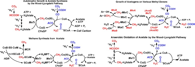
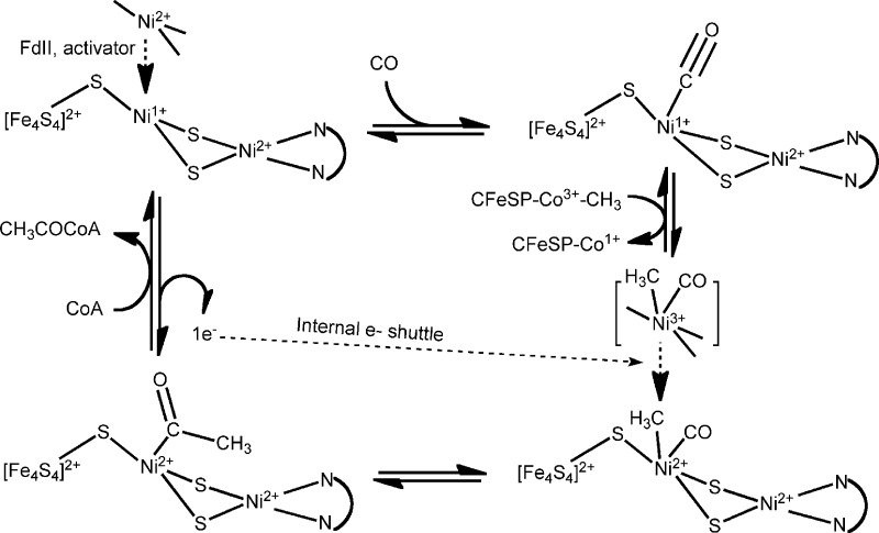
The Wood–Ljungdahl pathway of CO/CO2 fixation
and its involvement in acetogenesis and methyltrophy, as well as in
the oxidation of acetate to methane. The methanogenic CODH/ACS is
often called ACDS, acetyl-CoA synthase decarbonylase.
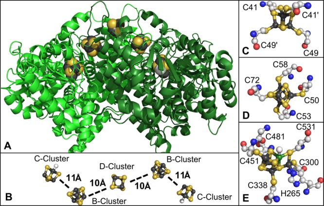
(A) Structure of CODHRr in cartoon
representation, (B) distances between the metal clusters, (C) structure
of the D-cluster, (D) structure of the B-cluster, and (E) structure
of the C-cluster. Atom colors: dark gray (iron), orange (sulfide),
red (oxygen), blue (nitrogen), white (carbon), dark green (nickel).
Generated using Pymol from PDB 1JQK.
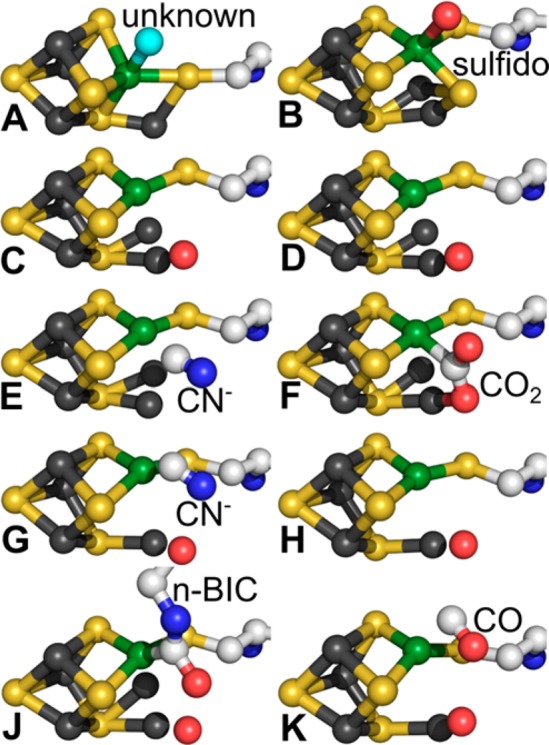
Structure of C-cluster including only one coordinating
residue,
cysteine, and the ligands from (A) CODHRr (PDB 1JQK),
(B) CODHCh II (PDB 1SU8), (C) CODHCh II at 320 mV (PDB 3B53), (D) CODHCh II at 600 mV (PDB 3B51), (E) cyanide-bound CODHCh II at 320
mV (PDB 3I39), (F) CO2-bound CODHCh II
at 600 mV (PDB 3B52), (G) cyanide-bound CODH/ACSMt (PDB 3I04), (H) CODH/ACSMt (PDB 3I01), (J) n-BICt-bound CODH/ACSMt (PDB 2YIV), (K) CO-bound CODHMb (PDB 3CF4). Atom colors: Dark gray (iron), orange (sulfide), red (oxygen),
blue (nitrogen), white (carbon), dark green (nickel).
The most well-characterized
ferredoxin (Fd) from M. thermoacetica and many other organisms contains two [Fe4S4] clusters and thus can accept two electrons. For a Fd containing
a single cluster, two Fd would be required.
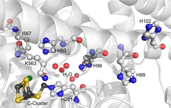
Structure of the C-cluster from CODHCh II at 600 mV including only one coordinating residue: histidine
and the ligands proposed to be important in catalytic activities.
Atom colors: Dark gray (iron), orange (sulfide), red (oxygen), blue
(nitrogen), white (carbon), dark green (nickel). Unbound red spheres
represent the water molecules. Generated using Pymol from PDB 3B51.
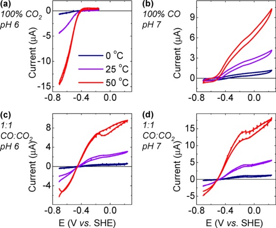
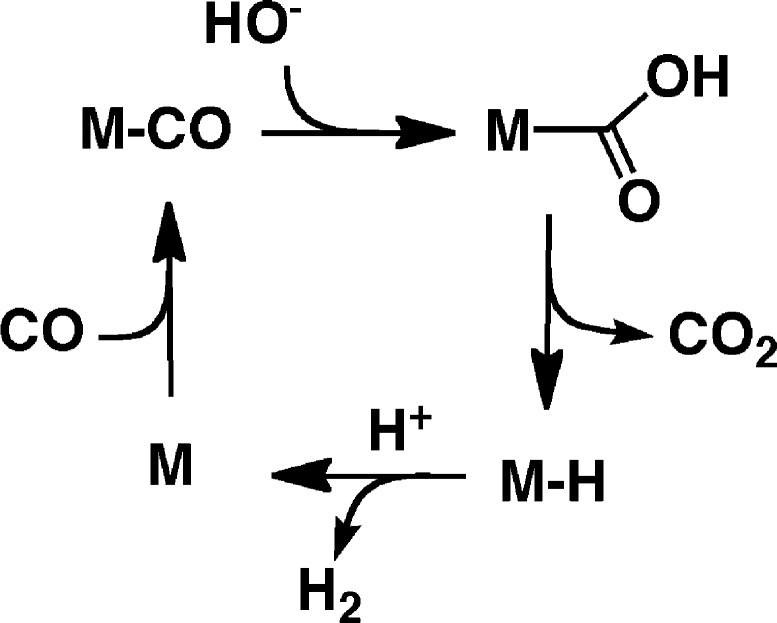
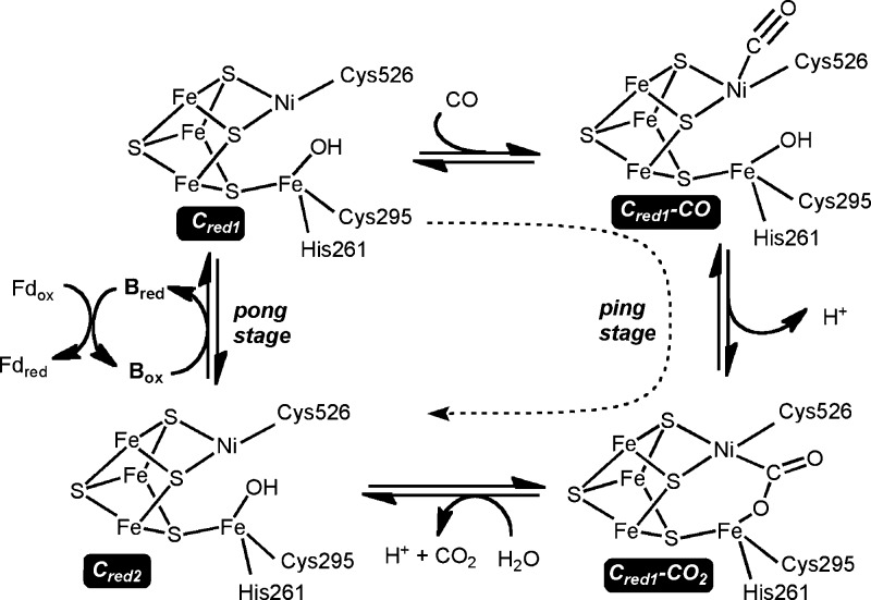
Protein film voltammograms showing CO2 reduction
and
CO oxidation activities of CODHCh I adsorbed
on a PGE electrode under atmospheres of 100% CO2, 100%
CO, or 1:1 CO2/CO gas mixtures. Scan rate was 10 mV/s in
parts a, c, and d and 30 mV/s in part b. Electrode rotation 4000 rpm.
Reprinted with permission from ref (45a). Copyright 2007 American Chemical Society.

Voltammograms showing, for CODHCh I,
(A) the potential dependence of inhibition of CO2 reduction
activity upon injection of cyanide (CO oxidation is completely inhibited),
pH 7.0, scan rate 1 mV s–1 and (B) inhibition of
CO2 reduction activity and shift in potential for CO oxidation
upon addition of cyanate, pH 7.0, scan rate 1 mV s–1. Adapted with permission from ref (45b). Copyright 2013 American Chemical Society.
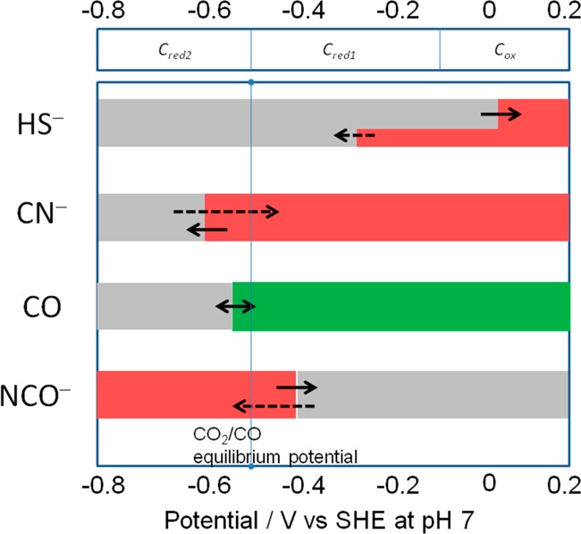
Potential dependence
of binding of inhibitors to CODHCh I.
Red refers to the potential region over which
the enzyme is inhibited, gray indicates no binding, and green indicates
that binding leads to turnover. The dashed arrows indicate reactions
that are slow compared to those indicated by full arrows. Reprinted
with permission from ref (45b). Copyright 2013 American Chemical Society.
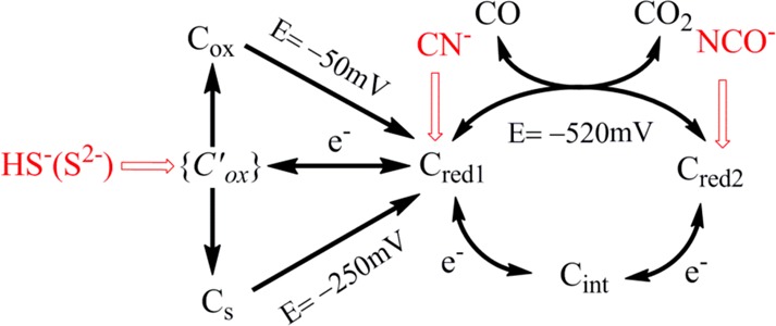
The potentials −250
and −50 mV are the values observed for reactivation of enzyme
with and without sulfide. The potential −520 mV is the standard
potential for the CO2/CO half-cell reaction at pH 7.0.
Reprinted with permission from ref (45b). Copyright 2013 American Chemical Society.
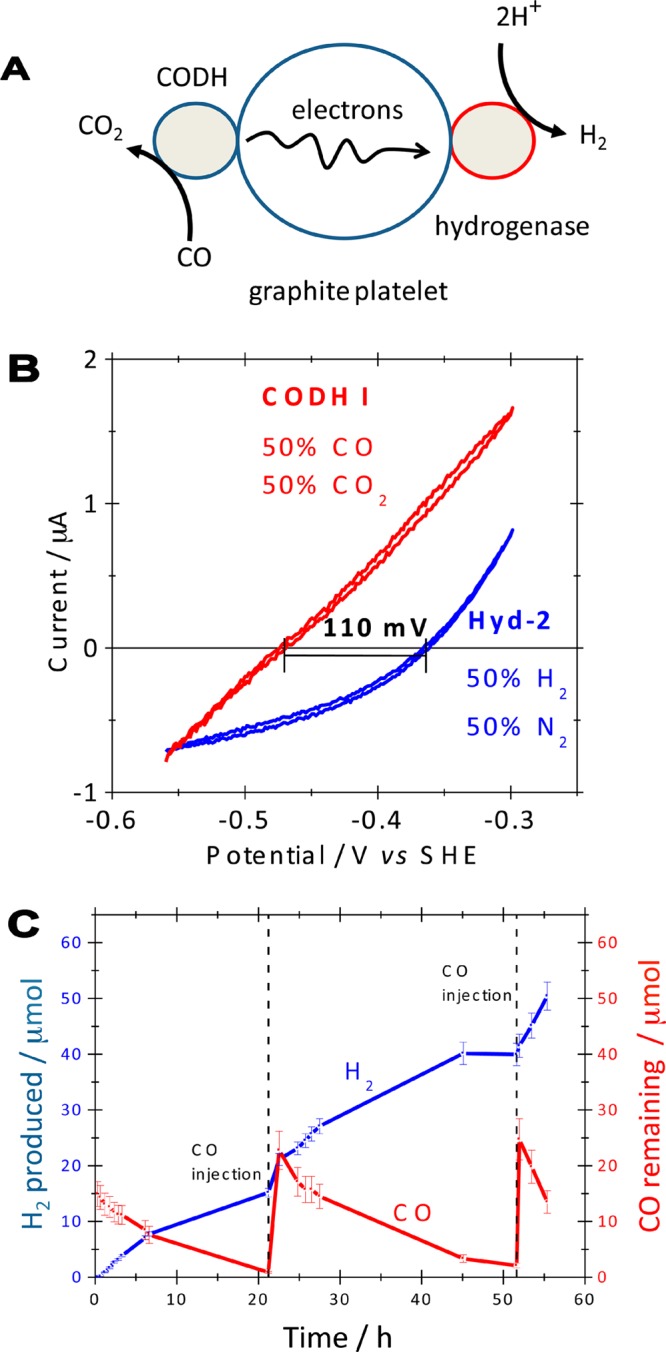
(A) Cartoon
representation of an enzymatic device for catalysis
of the water–gas shift reaction. Electrons released by CODH-catalyzed
CO oxidation are transferred through a graphite particle to a CO-tolerant
hydrogenase that reduces protons to H2. (B) Typical cyclic
voltammograms (from separate experiments) showing the reversibility
of electrocatalysis by CODHCh I and a
hydrogenase (Hyd-2) from E. coli, measured
at pH 6.0, 30 °C, scan rate 10 mV s–1, electrode
rotation rate 2500 rpm. (C) H2 production and CO depletion
over the course of 55 h at pH 6.0, 30 °C, as quantified by GC
analysis. Fresh aliquots of CO were introduced at the times indicated.
Adapted with permission from ref (51). Copyright 2009 American Chemical Society.
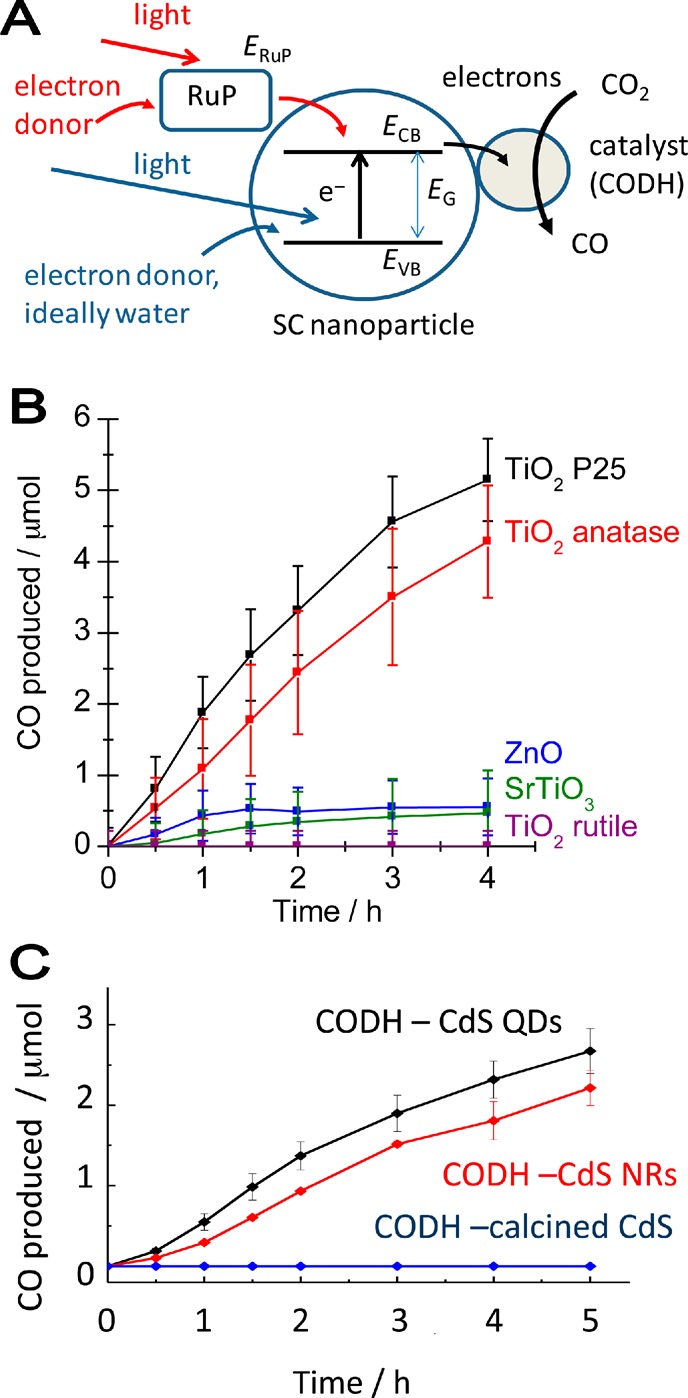
Photoelectrocatalysis of CO2 reduction to CO
catalyzed
by CODH attached to light-harvesting nanoparticles. (A) The concept:
red arrows correspond to injection of electron into the conduction
band (potential ECB) by a photosensitizer
(RuP) attached to the nanoparticle; green arrows correspond to injection
of electron into the conduction band by band gap excitation (potential
difference EG) from the valence band (potential EVB). The hole in either dye or valence band
must be filled more rapidly than the electron can return (the electron–hole
recombination rate). (B) Production of CO by visible light using a
photosensitizer. Experiments carried out by irradiating a vial containing
a 5 mL suspension of various semiconducting nanoparticles with visible
light (λ > 420 nm). In each case, 5 mg of nanoparticles (20
mg in the case of ZnO) was modified with CODHCh I (total 2.56 nmol) and RuP (total 56 nmol). The buffer in
each experiment was 0.20 M MES, pH 6, 20 °C. (C) Production of
CO by visible light using direct band gap excitation of various types
of cadmium sulfide attached to CODHCh I.
QD = quantum dot, NR = nanorod; calcined CdS was heated at 450 °C
for 45 min. The buffer in each experiment was 0.35 M MES, pH 6, at
20 °C. Adapted from refs (146a) (copyright 2011 The Royal Society of Chemistry)
and (147) (copyright
2012 The Royal Society of Chemistry) with permission.
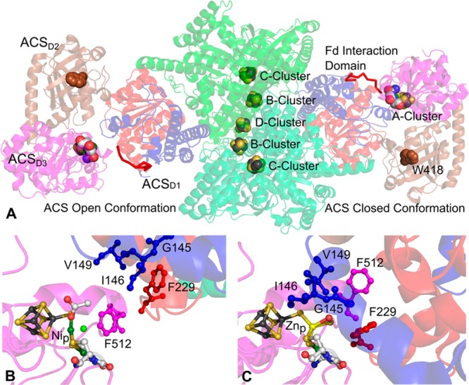
Structure
of CODH/ACSMt. (A) Overall
structure of CODH/ACS. Green units in the center are the two CODH
homodimers; the left unit is the ACS in open conformation, and the
right unit is the ACS in closed conformation. Closer views of the
A-cluster pocket in (B) open conformation and (C) closed conformation.
Atom colors: Brown (iron), orange (sulfide), red (oxygen), blue (nitrogen),
light green (carbon), dark green (nickel), white (unassigned). Generated
using Pymol from PDB 1OAO.
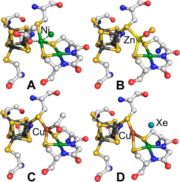
Structure of A-cluster from PDB (A and
B) 1OAO, (C) 1MJG, and (D) 2Z8Y. Generated using
Pymol.
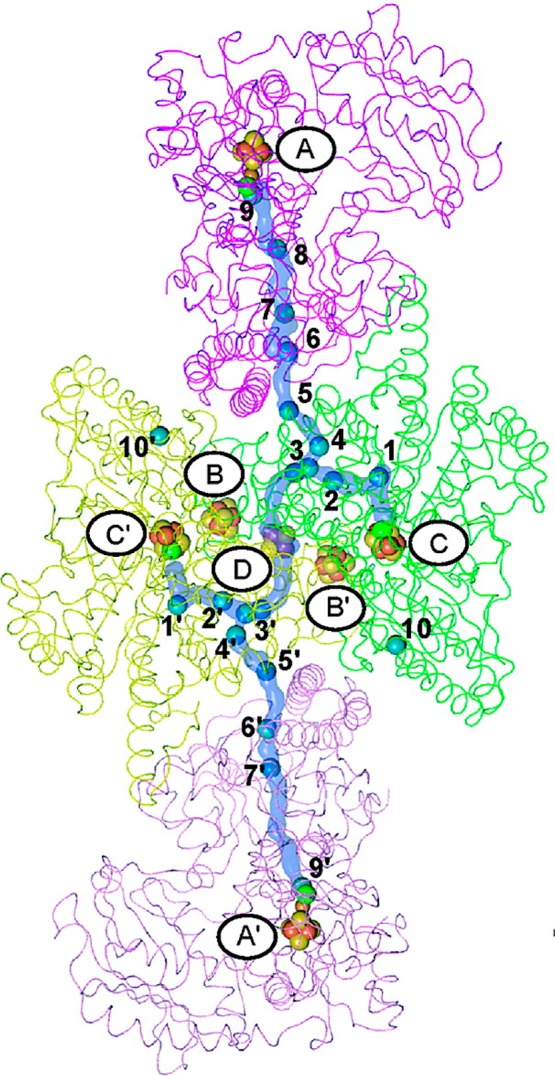
Structure of CODH/ACSMt crystallized
in the presence of high pressures of Xe (PDB 2Z8Y) (shown as the blue
spheres) to reveal the hydrophobic CO tunnel. Adapted with permission
from ref (71e). Copyright
2008 American Chemical Society.

References
-
- Yergin D.The Quest: Energy, Security and the Remaking of the Modern World; Penguin Press: New York, 2011.
-
- Hall D. O.; Rao K. K.. Photosynthesis, 5th ed.; Cambridge University Press: Cambridge, UK, 1994.
-
- Fuchs G. Annu. Rev. Microbiol. 2011, 65, 631. - PubMed
-
- Pollack H. N.A World without Ice; Avery/Penguin Group: New York, 2009.
Publication types
MeSH terms
Substances
Grants and funding
LinkOut - more resources
Full Text Sources
Other Literature Sources
Molecular Biology Databases
