. 2014 Feb 13;9(1):9.
doi: 10.1186/1749-799X-9-9.

Osteotomy at the distal third of tibial tuberosity with LCP L-buttress plate for correction of tibia vara

Affiliations

Osteotomy at the distal third of tibial tuberosity with LCP L-buttress plate for correction of tibia vara

Ye Huang et al. J Orthop Surg Res. .

Abstract

Background: Many osteotomy methods and fixation types have been used to correct the misalignment observed in tibia vara and to achieve a more uniform distribution of weight across the knee joint.

Purpose: The aim of this study is to test the efficacy and safety of a modified closing wedge high tibial osteotomy (CWHTO) procedure for tibia vara.

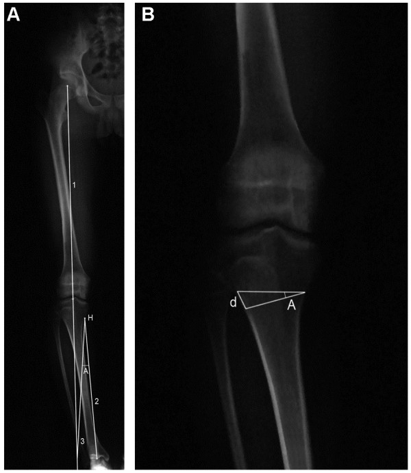
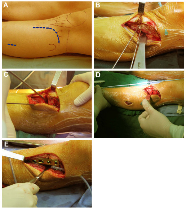
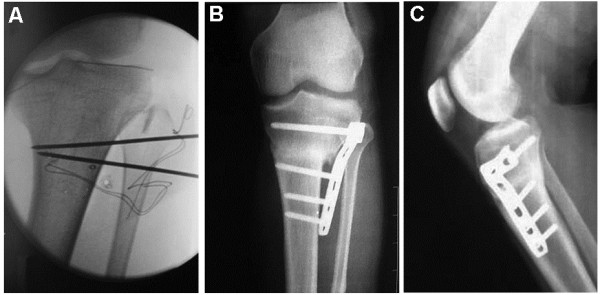
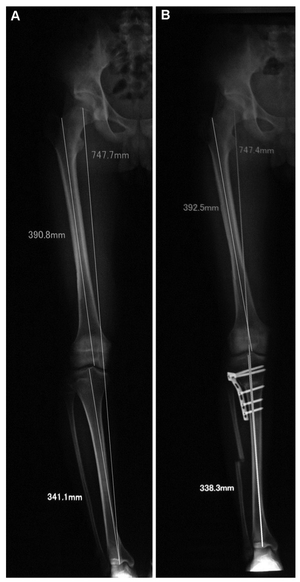
Methods: In this prospective study, young adults with tibia vara and mild medial arthritic changes were included. A CWHTO was performed at the distal third of the tibial tuberosity, instead of the conventional proximal site. An L-shaped locking compression plate was used for internal fixation. Before/after evaluation of femoro-tibial angle (FTA), pain relief, patellar height, and posterior tibial slope were evaluated. Adverse events were monitored.

Results: Seventy-five knees from 46 patients aged 27.2 ± 5.8 years (range, 14-43 years) underwent the modified CWHTO procedure. After a follow-up of 26.3 ± 7.4 months (range, 15-46 months), FTA correction was 12.4° ± 4.7° (range, 7°-31°), and pain was relieved. Reduction in the posterior tibial slope was 3.0° ± 2.3° (p<0.001), while there was no significant change in patella height. Bone union was observed in all patients. There were a delayed union in four knees, a peroneal nerve lesion in five knees causing partial paralysis and/or sensory loss, and infections in two knees. Three patients underwent a second surgery. All adverse events were successfully treated except for a case of extensor hallucis longus muscle paralysis.

Conclusions: The modified CWHTO procedure is efficient and safe for the correction of tibia vara in young patients.


Figures

Figure 1
Figure 1
Miniaci method for frontal plane correction. (A)Line 1 represents the planned Mikulicz line for the postoperative correction extending from the center of the hip through the center of the knee, past the ankle. Line 2 connects the osteotomy hinge point (H) with the center of the ankle. The H point was located on the medial cortex of the proximal tibia at the lower level of tibial tuberosity. With H point as the center and the length of line 2 as the radius, an angular arc is drawn from the center of the ankle to the intersection of line 1. Line 3 connects the H point with the intersection of line 1. The angle formed by lines 2 and 3 is the planned correction angle (A). (B)Angle A is now drawn on the prospective site of the osteotomy on the AP radiographs of the knee joint. The distance (d) of the two lines at the lateral cortex was measured as the osteotomy wedge base (magnification factor must be considered).
Figure 2
Figure 2
Surgical procedure for modified CWHTO. (A) A curvilinear incision was made over the lateral aspect of the proximal tibia, and a 3–4-cm incision was made over the lateral aspect of the leg at the level centered about 15 cm distal to fibular head. (B) The extensor muscles were detached to expose the lateral tibial cortex. The osteotomy site was at the distal third of the tibial tuberosity. Two K-wires were drilled in as osteotomy guides. (C) The cut was initiated with a saw under constant irrigation and completed with an osteotome. The hinge of the medial cortex was carefully penetrated with the use of a drill instead of an osteotome. (D) The wedge was closed and the frontal alignment was checked with a cautery cord. (E) An L-shaped LCP plate was inserted for internal fixation. The proximal part of the plate was placed at the level of Gerdy’s tubercle. The shaft of the plate was aligned to the longitudinal axis of the tibia. A full contact between the plate and the bone surface was not necessary.
Figure 3
Figure 3
Modified procedure for CWHTO. The osteotomy site was at the distal third of the tibial tuberosity. The procedure was performed under fluoroscopy, and an LCP was inserted for internal fixation. (A) Two K-wires were intraoperatively drilled in as osteotomy guides. The proximal wire was inserted at the level of the distal third of the tibial tuberosity and parallel to the joint line. The distal wire was then inserted in order to indicate the desired angle of correction and exited through the medial cortex where the first wire exited. The angle was determined from preoperative radiographic assessments and confirmed by fluoroscopy. (B, C) AP and lateral radiographs, respectively, 9 months after operation. The proximal fixation screws were at the level of or immediately distal to Gerdy’s tubercle, far from the articular surface. A full contact between the plate and bone surface was not necessary.
Figure 4
Figure 4
FTA correction by a modified CWHTO. (A) Preoperative; (B) 12 weeks postoperative. Full-length AP radiographs of a case of osteotomy showing correction of mechanical axis of lower limbs (Mikulicz line).

References

    1. McKellop HA, Llinas A, Sarmiento A. Effects of tibial malalignment on the knee and ankle. Orthop Clin North Am. 1994;25:415–423. - PubMed
    1. Jackson JP, Waugh W. Tibial osteotomy for osteoarthritis of the knee. J Bone Joint Surg Br. 1961;43-B:746–751. - PubMed
    1. Jackson JP, Waugh W. The technique and complications of upper tibial osteotomy. A review of 226 operations. J Bone Joint Surg Br. 1974;56:236–245. - PubMed
    1. Coventry MB. Osteotomy of the upper portion of the tibia for degenerative arthritis of the knee. A preliminary report. J Bone Joint Surg Am. 1965;47:984–990. - PubMed
    1. Moreland JR, Bassett LW, Hanker GJ. Radiographic analysis of the axial alignment of the lower extremity. J Bone Joint Surg Am. 1987;69:745–749. - PubMed

Supplementary concepts

LinkOut - more resources