. 2014 Jul;24(4):360-70.
doi: 10.1111/bpa.12131. Epub 2014 Mar 28.

Cripto-1 expression in glioblastoma multiforme

Affiliations

Cripto-1 expression in glioblastoma multiforme

Linda Pilgaard et al. Brain Pathol. 2014 Jul.

Abstract

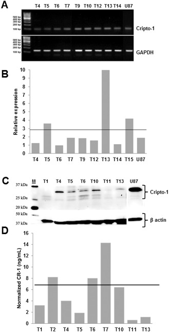
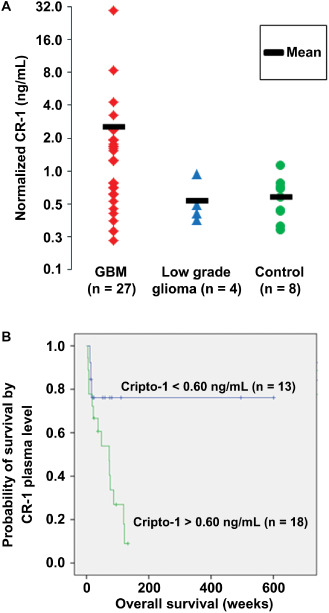
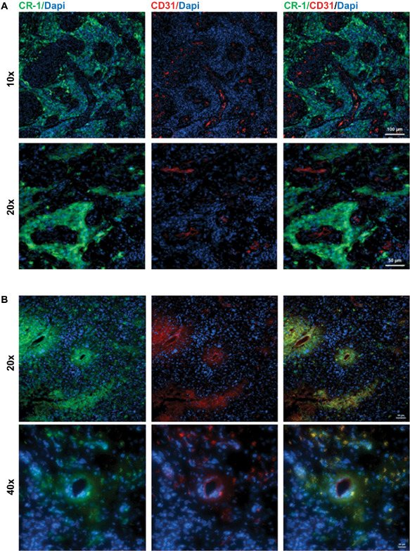
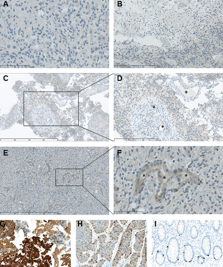
Human glioblastoma multiforme (GBM) is an aggressive cancer with a very poor prognosis. Cripto-1 (CR-1) has a key regulatory role in embryogenesis, while in adult tissue re-expression of CR-1 has been correlated to malignant progression in solid cancers of non-neuronal origin. As CR-1 expression has yet to be described in cerebral cancer and CR-1 is regulated by signaling pathways dysregulated in GBM, we aimed to investigate CR-1 in the context of expression in GBM. The study was performed using enzyme-linked immunosorbent assay (ELISA), Western blotting, polymerase chain reaction (PCR) and immunohistochemistry to analyze the blood and tissue from 28 GBM and 4 low-grade glioma patients. Within the patient cohort, we found high CR-1 protein levels in blood plasma to significantly correlate with a shorter overall survival. We identified CR-1 in different areas of GBM tissue, including perivascular tumor cells, and in endothelial cells. Collectively, our data suggest that CR-1 could be a prognostic biomarker for GBM with the potential of being a therapeutic target.

Keywords: CR-1; endothelial proliferation; glioblastoma multiforme; microvasculature; plasma biomarker; tumor niche.


Figures

Figure 1
Figure 1
Presence of Cripto‐1 ( CR ‐1) in glioblastoma multiforme ( GBM ) tumor tissue. A. CR‐1 messenger RNA (mRNA) by reverse transcription polymerase chain reaction (RTPCR) in GBM tumor samples. B. CR‐1 mRNA by quantitative RTPCR in GBM tumor samples. Normalization was performed relative to hypoxanthine‐guanine phosphoribosyltransferase (HPRT). Horizontal line depicts the mean relative expression for GBM samples of 2.9. C. CR‐1 protein in GBM tumor samples by Western blotting. D. CR‐1 protein in GBM tumor samples quantified by enzyme‐linked immunosorbent assay (ELISA). Horizontal line represents the mean normalized concentration of 5.3 ng/mL. Normalization is performed relative to the total protein contents of each sample. Tx denotes the tumor samples of individual patients.
Figure 2
Figure 2
Cripto‐1 ( CR ‐1) in glioma patient blood and its association with overall survival of the patients. A. CR‐1 protein in glioblastoma multiforme (GBM) plasma samples, normal control plasma and plasma of lower grade glioma patient. Normalization is performed relative to the total protein contents of each sample. B. Kaplan–Meier plot showing overall survival in glioblastoma and low‐grade glioma patients by plasma levels of CR‐1 above or below the average control level set at 0.60 ng/mL. Symbols on the line indicate patients alive at the time of analysis.
Figure 3
Figure 3
Co‐localization of Cripto‐1 ( CR ‐1) withCD 31 in patientsT 24 andT 25. A. CR‐1‐positive cells present in the niche surrounding glomeruloid vasculature but not co‐expressed with the endothelial marker CD31 in patient T24. Scale bar indicates 100 μm (top panel) and 50 μm (lower panel). B. Areas containing microvasculature with CR‐1‐positive cells co‐localizing with CD31 expressing endothelial cells lining capillaries in patient T25. Scale bar indicates 50 μm (top panel) and 20 μm (lower panel).
Figure 4
Figure 4
Immunohistochemical staining for Cripto‐1 ( CR ‐1) in glioblastoma tissue from patients presentingCR ‐1 low and high plasma levels, respectively. A. CR‐1 staining in CR‐1 low patient T11. Barely perceptible staining in endothelial cells (left). B. Faint CR‐1 staining in pseudopalisading cells around necrotic area of low cell density in patient T6. C,D. Faint CR‐1 staining in the vicinity of glomeruloid vasculature in CR‐1 high patient T7. E,F. Moderate CR‐1 staining in endothelial cells in CR‐1 high patient T6. G. CR‐1 staining of formalin‐fixed, paraffin‐embedded U87 cerebral xenograft showing strong and medium mainly cytoplasmic reaction in tumor cells and no staining in murine neural cells. H. CR‐1 staining of small intestine neuroendocrine tumor (carcinoid) showing a distinct, heterogenous cytoplasmic staining. I. CR‐1 staining of colon mucosa showing strong cytoplasmic staining of neuroendocrine crypt cells. *Vessel lumen. Scale bar indicates 800 μm (C and E), 300 μm (B and D), 100 μm (A and F) and 200 μm (G, H and I).

References

    1. Adkins HB, Bianco C, Schiffer SG, Rayhorn P, Zafari M, Cheung AE et al (2003) Antibody blockade of the Cripto CFC domain suppresses tumor cell growth in vivo . J Clin Invest 112:575–587. - PMC - PubMed
    1. Beier D, Hau P, Proescholdt M, Lohmeier A, Wischhusen J, Oefner PJ et al (2007) CD133(+) and CD133(−) glioblastoma‐derived cancer stem cells show differential growth characteristics and molecular profiles. Cancer Res 67:4010–4015. - PubMed
    1. Bianco C, Castro NP, Baraty C, Rollman K, Held N, Rangel MC et al (2013) Regulation of human Cripto‐1 expression by nuclear receptors and DNA promoter methylation in human embryonal and breast cancer cells. J Cell Physiol 228:1174–1188. - PMC - PubMed
    1. Bianco C, Cotten C, Lonardo E, Strizzi L, Baraty C, Mancino M et al (2009) Cripto‐1 is required for hypoxia to induce cardiac differentiation of mouse embryonic stem cells. Am J Pathol 175:2146–2158. - PMC - PubMed
    1. Bianco C, Rangel MC, Castro NP, Nagaoka T, Rollman K, Gonzales M, Salomon DS (2010) Role of Cripto‐1 in stem cell maintenance and malignant progression. Am J Pathol 177:532–540. - PMC - PubMed

Publication types

MeSH terms

Substances