. 2014 Apr;21(4):534-45.
doi: 10.1128/CVI.00688-13. Epub 2014 Feb 12.

Trained immunity or tolerance: opposing functional programs induced in human monocytes after engagement of various pattern recognition receptors

Affiliations

Trained immunity or tolerance: opposing functional programs induced in human monocytes after engagement of various pattern recognition receptors

Daniela C Ifrim et al. Clin Vaccine Immunol. 2014 Apr.

Abstract

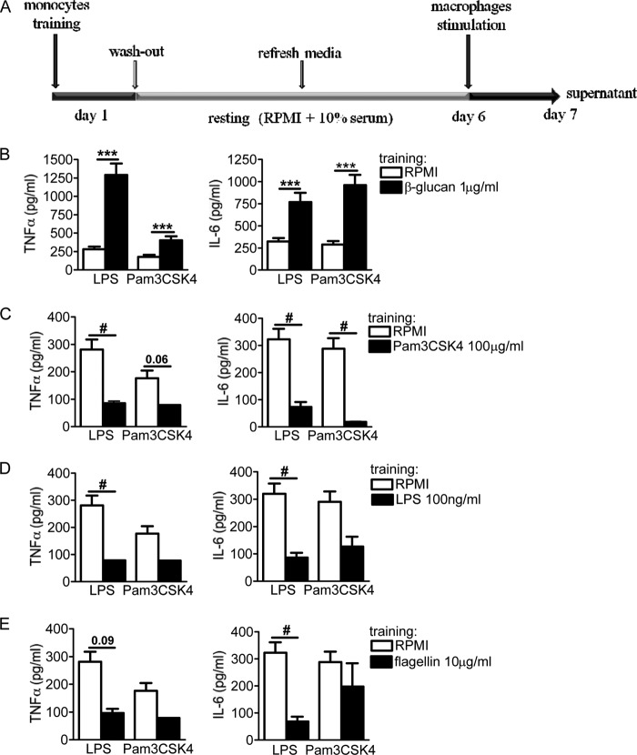
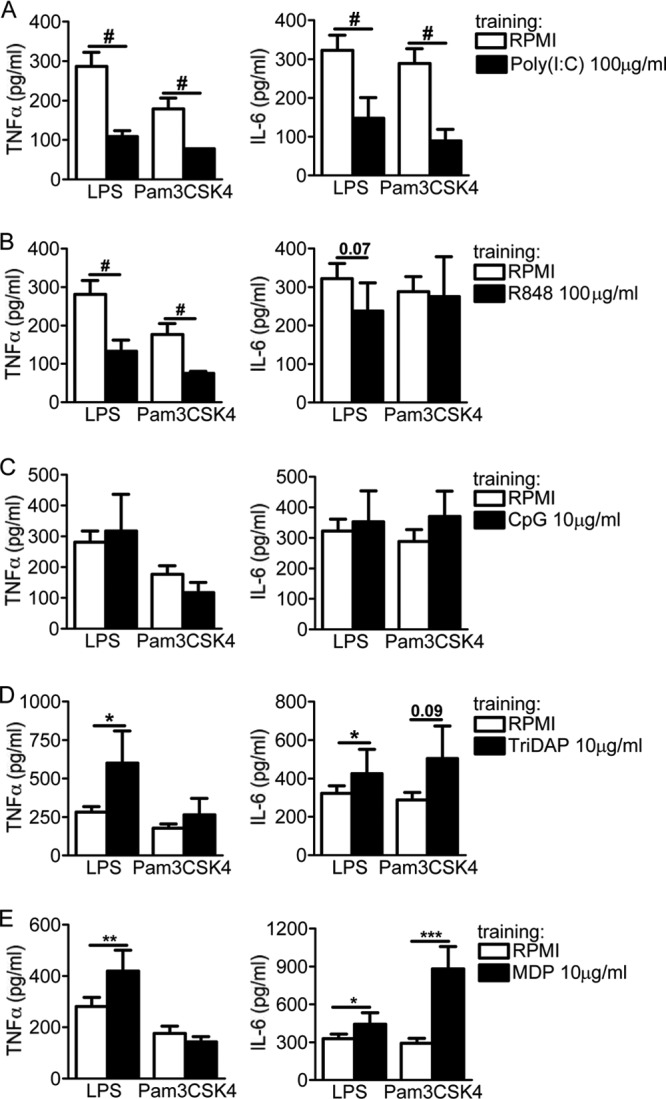
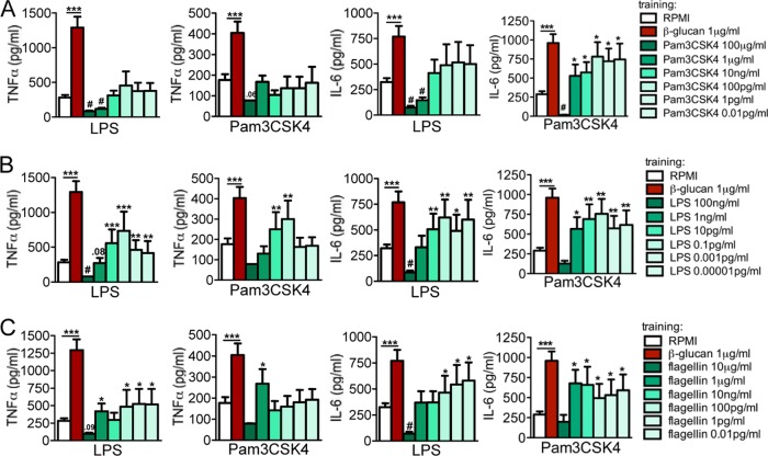
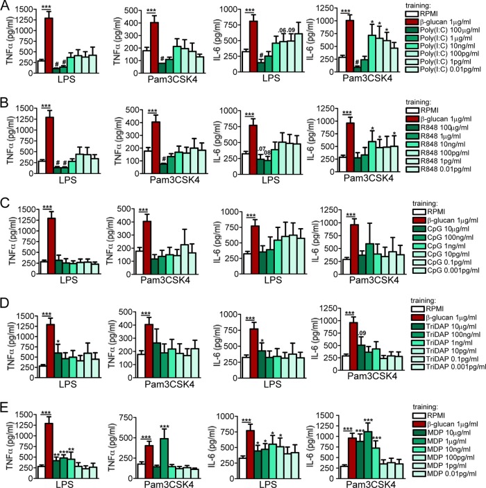
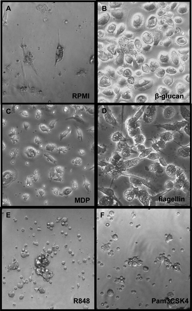
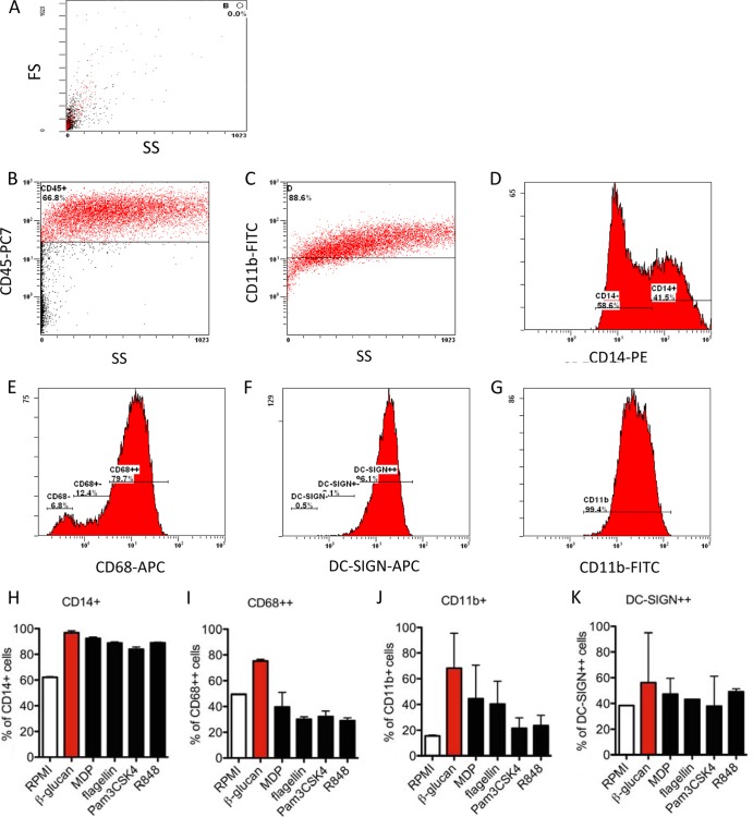
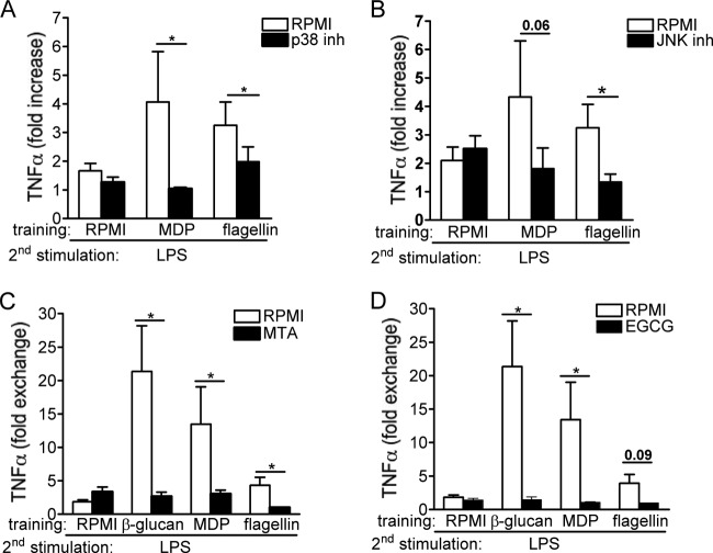
Upon priming with Candida albicans or with the fungal cell wall component β-glucan, monocytes respond with an increased cytokine production upon restimulation, a phenomenon termed "trained immunity." In contrast, the prestimulation of monocytes with lipopolysaccharide has long been known to induce tolerance. Because the vast majority of commensal microorganisms belong to bacterial or viral phyla, we sought to systematically investigate the functional reprogramming of monocytes induced by the stimulation of pattern recognition receptors (PRRs) with various bacterial or viral ligands. Monocytes were functionally programmed for either enhanced (training) or decreased (tolerance) cytokine production, depending on the type and concentration of ligand they encountered. The functional reprogramming of monocytes was also associated with cell shape, granulocity, and cell surface marker modifications. The training effect required p38- and Jun N-terminal protein kinase (JNK)-mediated mitogen-activated protein kinase (MAPK) signaling, with specific signaling patterns directing the functional fate of the cell. The long-term effects on the function of monocytes were mediated by epigenetic events, with both histone methylation and acetylation inhibitors blocking the training effects. In conclusion, our experiments identify the ability of monocytes to acquire adaptive characteristics after prior activation with a wide variety of ligands. Trained immunity and tolerance are two distinct and opposing functional programs induced by the specific microbial ligands engaging the monocytes.


Figures

FIG 1
FIG 1
(A) In vitro training scheme. Adherent monocytes were incubated with pure ligands (training stimuli) for 24 h at 37°C. Following the first incubation, the cells were washed with warm PBS and further incubated with RPMI and pooled human serum for 5 days. In the absence of any training stimulus during the first 24 h, primary monocytes maintained in culture for several days eventually differentiated into macrophages. After this resting period, different pattern recognition receptor ligands were added (second stimulus) for an additional 24 h. (B to E) Tolerance induced by the membrane receptors. The cells were preexposed for 24 h to culture medium, β-glucan (1 μg/ml) (B), Pam3CSK4 (100 μg/ml) (C), LPS (100 ng/ml) (D), or flagellin (10 μg/ml) (E). Primary stimuli were washed out, and after 6 days, the macrophages were restimulated with RPMI (negative control), LPS (10 ng/ml), or Pam3CSK4 (10 μg/ml). While β-glucan induced training, Pam3CSK4, LPS, and flagellin induced tolerance. The data are presented as means ± standard error of the mean (SEM) (n > 8, from 5 independent experiments). *, P < 0.05; **, P < 0.01; ***, P < 0.001; #, P < 0.05 (tolerance). The Wilcoxon signed-rank test was used to detect significant differences.
FIG 2
FIG 2
Differential effects of endosomal and cytosolic receptors on adherent monocytes. The cells were preexposed for 24 h to culture medium, poly(I·C) (100 μg/ml) (A), R848 (100 μg/ml) (B), CpG (10 μg/ml) (C), Tri-DAP (10 μg/ml) (D), and MDP (10 μg/ml) (E). Primary stimuli were washed out, and after 6 days, the macrophages were restimulated with RPMI (negative control), LPS (10 ng/ml), or Pam3CSK4 (10 μg/ml). While poly(I·C) induced tolerance for TNF-α and IL-6, R848 was able to induce tolerance for TNF-α only, and CpG did not exert any significant effect on the inflammatory status of monocytes. Both types of NLRs were able to enhance the status of monocytes upon a second exposure to either LPS or Pam3CSK4. The data are presented as the means ± SEM (n > 8, from 5 independent experiments). *, P < 0.05; **, P < 0.01; ***, P < 0.001; #, P < 0.05 (tolerance). The Wilcoxon signed-rank test was used to detect significant differences.
FIG 3
FIG 3
Dose responses of membrane-bound receptors. The cells were preexposed for 24 h to culture medium or to different concentrations of Pam3CSK4 (100 μg/ml to 0.01 pg/ml) (A), LPS (100 ng/ml to 10−5 pg/ml) (B), and flagellin (10 μg/ml to 0.01 pg/ml) (C). The first stimuli were washed away, and adherent monocytes were further incubated for 5 days in culture medium supplemented with 10% pooled human pool serum. At day 6, the cells were restimulated with RPMI (negative control), LPS (10 ng/ml), or Pam3CKS4 (10 μg/ml). Depending on the concentration used, after several days, cells entered either a trained or tolerized status. The data are presented as the means ± SEM (n > 8, from 5 independent experiments), *, P < 0.05; **, P < 0.01; ***, P < 0.001; #, P < 0.05 (tolerance). The Wilcoxon signed-rank test was used to detect significant differences. TNF-α and IL-6 from cells stimulated with RPMI were below the detection limit.
FIG 4
FIG 4
Dose responses of endosomal and cytosolic receptors. The cells were preexposed for 24 h to culture medium or to different concentrations of poly(I·C) (100 μg/ml to 0.01 pg/ml) (A), R848 (100 μg/ml to 0.01 pg/ml) (B), CpG (10 μg/ml to 0.001 pg/ml) (C), Tri-DAP (10 μg/ml to 0.001 pg/ml) (D), and MDP (10 μg/ml to 0.01 pg/ml) (E). The first stimuli were washed away, and adherent monocytes were further incubated for 5 days in culture medium supplemented with 10% pooled human pool serum. At day 6, the cells were restimulated with RPMI (negative control), LPS (10 ng/ml), or Pam3CKS4 (10 μg/ml). Depending on concentration used, after several days, cells entered either a trained or tolerized status. The data are presented as the means ± SEM (n > 8, from 5 independent experiments), *, P < 0.05; **, P < 0.01; ***, P < 0.001; #, P < 0.05 (tolerance). Wilcoxon signed-rank test was used to detect significant differences. TNF-α and IL-6 from cells stimulated with RPMI were below the detection limit.
FIG 5
FIG 5
Morphology of trained versus tolerized cells. The cells were preexposed for 24 h to culture medium (A) or to several pure ligands, such as β-glucan (1 μg/ml) (B), MDP (1 μg/ml) (C), flagellin (1 μg/ml) (D), R848 (1 μg/ml) (E), and Pam3CSK4 (1 μg/ml) (F). The first stimuli were washed away, and adherent monocytes were further incubated for 5 days in culture medium supplemented with 10% pooled human pool serum. At day 6, the cells were microscopically examined and representative pictures were chosen (×200). After several days in culture, primary monocytes differentiated into the macrophages. While trained cells appeared to be bigger and more activated (B, to D), cells that entered a tolerizing status seemed to be considerably smaller (E and F).
FIG 6
FIG 6
Flow cytometric analysis of CD14-, CD68-, DC-SIGN-, and CD11b-positive cells gated on CD45+ populations after prestimulation with β-glucan, MDP, flagellin, R848, and Pam3CSK4. A total of 3 ml at 5 × 10⁶ PBMCs was incubated for 1 h, washed 3 times with PBS, and the remaining adherent monocytes were coincubated with β-glucan (1 μg/ml), MDP (1 μg/ml), flagellin (1 μg/ml), R848 (1 μg/ml), and Pam3CSK4 (1 μg/ml), or with RPMI (untrained control) for 24 h. Thereafter, the cells were washed and further incubated with RPMI supplemented with 10% pooled human serum for 5 days. At day 6, the macrophages were incubated with Versene solution for 1 h at 37°C, detached from the plate, and stained extracellularly using anti-CD45 PECy7, anti-CD14 PE, anti-CD68 APC, anti-DC-SIGN APC, and anti-CD11b FITC antibody. (A) The side scatter (SS) and forward scatter (FS) of the RPMI-treated cells are shown. (B and C) One representative picture of the staining and gating on CD45+ high β-glucan-trained cells is shown. Depending on the fluorescence intensity as well as cell size, we identified several cell populations, depicted as negative (−), positive (+), and high positive (++). Gating on different populations of CD14+ β-glucan-trained cells (D), CD68+ β-glucan-trained cells (E), DC-SIGN+ β-glucan-trained cells (F), and CD11b+ β-glucan-trained cells (G) are shown. (H to K) % of positive cells gated on CD45+ high. The data are presented as the means ± SEM (n > 2, from 4 independent experiments).
FIG 7
FIG 7
(A and B) MAP kinase-dependent pathways mediate trained immunity. Adherent monocytes were preincubated for 1 h with culture medium, p38, ERK, JNK, or Syk inhibitor (inh). After this inhibition period, 100 μl per well of either culture medium, flagellin (1 μg/ml), or MPD (1 μg/ml) was added to the cell suspension, without removing the inhibitors, for 24 h. Thereafter, the cells were washed and incubated with RPMI and 10% pooled human serum. At day 6, the cells were subjected to a second stimulation with LPS (10 ng/ml) for an additional 24 h. The data are presented as the means ± SEM (n ≥ 7, from four independent experiments). *, P <0.05. The Wilcoxon signed-rank test was used to detect significant differences. (C and D) Blocking histone methylation and acetylation inhibits trained immunity induced by MDP and flagellin. Before training, adherent monocytes were preincubated for 1 h with culture medium or with different epigenetic inhibitors, such as histone methyltransferase inhibitor (MTA), histone acetyltransferase inhibitor (EGCG), histone demethylase inhibitor (pargyline), and the histone deacetylase inhibitor ITF2357/9. Thereafter, 100 μl of culture medium, β-glucan (10 μg/ml), flagellin (1 μg/ml), or MPD (1 μg/ml) was added to the cell suspension, without removing the inhibitors, and further incubated for an additional 24 h. At day 6, the macrophages were subjected to a second stimulation with LPS (10 ng/ml) for another 24 h. The data are presented as the means ± SEM (n = 6, from three independent experiments), *, P < 0.05. The Wilcoxon signed-rank test was used to detect significant differences.

References

    1. Faulhaber LM, Karp RD. 1992. A diphasic immune response against bacteria in the American cockroach. Immunology 75:378–381 - PMC - PubMed
    1. Rheins LA, Karp RD, Butz A. 1978. Humoral Immunity Induced in American Cockroach (Periplaneta americana). Am. Zool. 18:595
    1. Rheins LA, Karp RD. 1984. The humoral immune response in the American cockroach, Periplaneta americana: reactivity to a defined antigen from honeybee venom, phospholipase A2. Dev. Comp. Immunol. 8:791–801. 10.1016/0145-305X(84)90062-4 - DOI - PubMed
    1. Sequeira T, Tavares D, Arala-Chaves M. 1996. Evidence for circulating hemocyte proliferation in the shrimp Penaeus japonicus. Dev. Comp. Immunol. 20:97–104. 10.1016/0145-305X(96)00001-8 - DOI - PubMed
    1. Arala-Chaves M, Sequeira T. 2000. Is there any kind of adaptive immunity in invertebrates? Aquaculture 191:247–258. 10.1016/S0044-8486(00)00430-0 - DOI

Publication types