

Google Glass for documentation of medical findings: evaluation in forensic medicine
- PMID: 24521935
- PMCID: PMC3936278
- DOI: 10.2196/jmir.3225
Google Glass for documentation of medical findings: evaluation in forensic medicine
Abstract
Background: Google Glass is a promising premarket device that includes an optical head-mounted display. Several proof of concept reports exist, but there is little scientific evidence regarding its use in a medical setting.
Objective: The objective of this study was to empirically determine the feasibility of deploying Glass in a forensics setting.
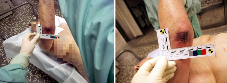
Methods: Glass was used in combination with a self-developed app that allowed for hands-free operation during autopsy and postmortem examinations of 4 decedents performed by 2 physicians. A digital single-lens reflex (DSLR) camera was used for image comparison. In addition, 6 forensic examiners (3 male, 3 female; age range 23-48 years, age mean 32.8 years, SD 9.6; mean work experience 6.2 years, SD 8.5) were asked to evaluate 159 images for image quality on a 5-point Likert scale, specifically color discrimination, brightness, sharpness, and their satisfaction with the acquired region of interest. Statistical evaluations were performed to determine how Glass compares with conventionally acquired digital images.
Results: All images received good (median 4) and very good ratings (median 5) for all 4 categories. Autopsy images taken by Glass (n=32) received significantly lower ratings than those acquired by DSLR camera (n=17) (region of interest: z=-5.154, P<.001; sharpness: z=-7.898, P<.001; color: z=-4.407, P<.001, brightness: z=-3.187, P=.001). For 110 images of postmortem examinations (Glass: n=54, DSLR camera: n=56), ratings for region of interest (z=-8.390, P<.001) and brightness (z=-540, P=.007) were significantly lower. For interrater reliability, intraclass correlation (ICC) values were good for autopsy (ICC=.723, 95% CI .667-.771, P<.001) and postmortem examination (ICC=.758, 95% CI .727-.787, P<.001). Postmortem examinations performed using Glass took 42.6 seconds longer than those done with the DSLR camera (z=-2.100, P=.04 using Wilcoxon signed rank test). The battery charge of Glass quickly decreased; an average 5.5% (SD 1.85) of its battery capacity was spent per postmortem examination (0.81% per minute or 0.79% per picture).
Conclusions: Glass was efficient for acquiring images for documentation in forensic medicine, but the image quality was inferior compared to a DSLR camera. Images taken with Glass received significantly lower ratings for all 4 categories in an autopsy setting and for region of interest and brightness in postmortem examination. The effort necessary for achieving the objectives was higher when using the device compared to the DSLR camera thus extending the postmortem examination duration. Its relative high power consumption and low battery capacity is also a disadvantage. At the current stage of development, Glass may be an adequate tool for education. For deployment in clinical care, issues such as hygiene, data protection, and privacy need to be addressed and are currently limiting chances for professional use.
Keywords: Google Glass; autopsy, postmortem examination; documentation; forensic medicine.
Conflict of interest statement
Conflicts of Interest: None declared.
Figures


References
-
- Dexheimer JW, Borycki EM. Use of mobile devices in the emergency department. Stud Health Technol Inform. 2013;192:1086. - PubMed
-
- Google Glass. 2014. [2014-01-03]. http://www.google.com/glass/start/
-
- Google. 2014. [2014-01-03]. The Glass Explorer Program http://www.google.com/glass/start/how-to-get-one/
Publication types
MeSH terms
LinkOut - more resources
Full Text Sources
Other Literature Sources
