. 2014 May;15(5):524-32.
doi: 10.4161/cbt.28018. Epub 2014 Feb 12.

Caffeine and the analog CGS 15943 inhibit cancer cell growth by targeting the phosphoinositide 3-kinase/Akt pathway

Affiliations

Caffeine and the analog CGS 15943 inhibit cancer cell growth by targeting the phosphoinositide 3-kinase/Akt pathway

Charlotte E Edling et al. Cancer Biol Ther. 2014 May.

Abstract

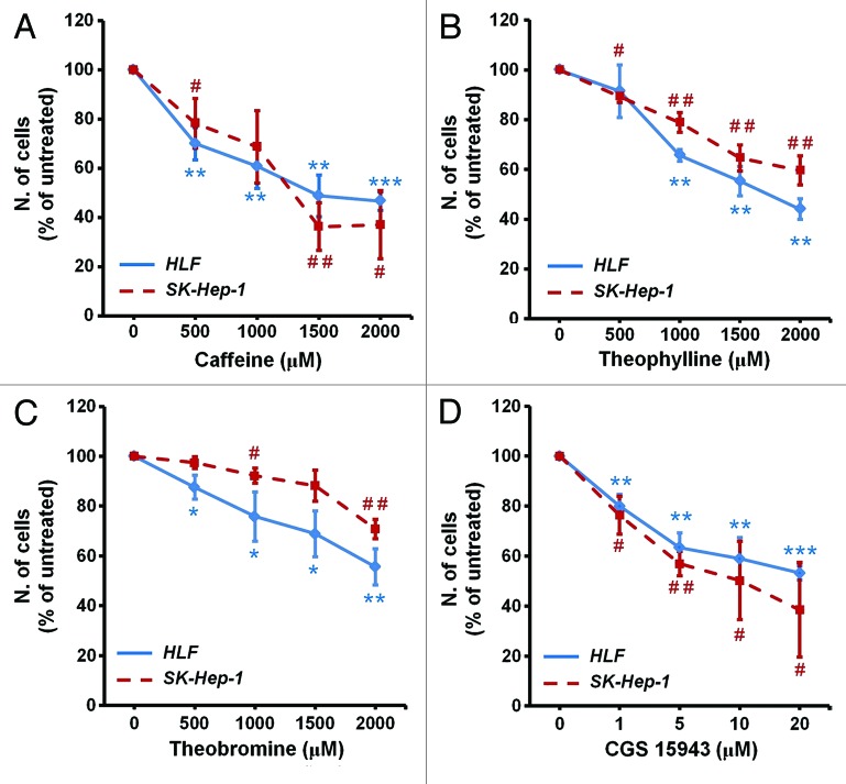
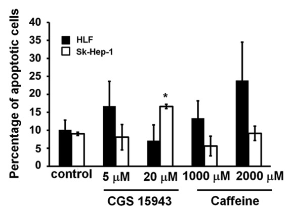
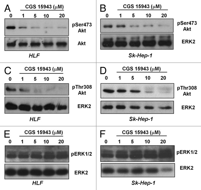
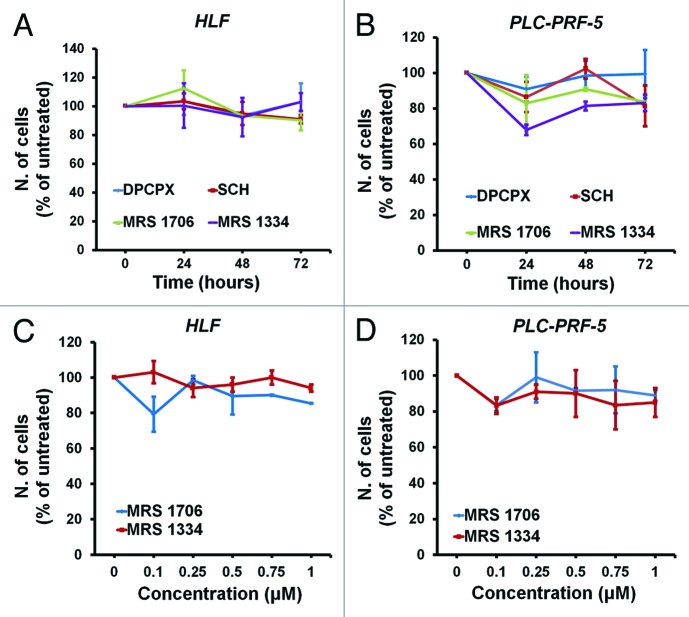
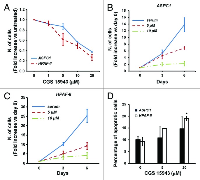
Caffeine is a naturally occurring methylxanthine that acts as a non-selective adenosine receptor antagonist. Epidemiological studies demonstrated habitual coffee drinking to be significantly associated with liver cancer survival. We aimed to investigate the effects of caffeine and its analog CGS 15943 on hepatocellular carcinoma (HCC) and pancreatic cancer adenocarcinoma (PDAC). We demonstrate that caffeine and CGS 15943 block proliferation in HCC and PDAC cell lines by inhibiting the PI3K/Akt pathway. Importantly a kinase profiling assay reveals that CGS 15943 targets specifically the catalytic subunit of the class IB PI3K isoform (p110γ). These data give mechanistic insight into the action of caffeine and its analogs and they identify these compounds as promising lead compounds to develop drugs that can specifically target this PI3K isoform whose key role in cancer progression is emerging.

Keywords: CGS 15943; caffeine; hepatocellular carcinoma; pancreatic ductal adenocarcinoma; phosphoinositide 3-kinase.


Figures

None
Figure 1. In vitro activity of xanthines and CGS 15943 on HCC cell lines. HLF and SK-Hep-1 cell lines were treated for 72 h with increasing concentrations of the indicated compounds in the presence of serum and cell proliferation was assessed by cell counting. In all panels data are expressed as percentage of untreated cells and are means ± s.e.m. (A) Results are from n = 4–7 (HLF) and n = 3–5 (Sk-Hep-1) independent experiments. (B) Results are from n = 3 (HLF) and n = 4 (Sk-Hep-1) independent experiments. (C) For both cell lines results are from n = 4 independent experiments. (D) Results are from n = 4–7 (HLF) and n = 3–4 (Sk-Hep-1) independent experiments. HLF: *P < 0.05, **P < 0.01, ***P < 0.001 vs corresponding untreated cells as assessed by the Student t test (paired, one-tailed distribution). SK-Hep-1: #P < 0.05, ##P < 0.01 vs corresponding untreated cells as assessed by the Student t test (paired, one-tailed distribution).
None
Figure 2. Anti-apoptotic activity of CGS 154943 and caffeine in HCC cells. HCC cell lines were treated for 72 h with the indicated concentrations of CGS 15943 and caffeine. The number of surviving cells was assessed by flow cytometry using Annexin V-conjugated FITC and PI stain. Data are means ± s.e.m. of n = 3 (SK-Hep-1) and n = 2 (HLF) independent experiments. *P < 0.01.
None
Figure 3. In vitro activity of CGS and caffeine on Akt phosphorylation. HLF and SK-Hep-1 cells were treated for 24 h with the indicated concentrations of CGS 15943 in the presence of serum. Akt activation was assessed by monitoring phosphorylation at its residues Ser473 (A and B) and Thr308 (C and D). Equal loading was assessed using an anti-ERK2 antibody. Alternatively membranes were stripped and re-incubated with an anti-Akt antibody. Phosphorylation of ERK1/2 was also assessed by using a specific antibody (E and F). Membranes were then stripped and re-incubated with an anti-ERK2 antibody.
None
Figure 4. In vitro activity of adenosine receptors-specific antagonists on HCC cell lines. HLF and PLC-PRF-5 cells were treated for the indicated time points with 0.1 μM of the indicated adenosine receptors antagonists (A and B) or for 72 h with increasing concentrations of receptors-specific antagonists (C and D) in the presence of serum. Cell growth was assessed by cell counting. In all panels data are expressed as percentage of untreated cells and are means ± s.e.m. of n = 2 (A and C) (except for treatment with MRS 1796 and MRS 1334 at 72 h for which n = 4) and n = 2 (B and D) (except for treatment at 0.1 μM for which n = 4) independent experiments.
None
Figure 5. In vitro activity of caffeine and CGS 15943 on PDAC cell lines. (A) ASPC1 and HPAF-II cells were treated for 72 h with the indicated concentrations of caffeine and CGS 15943 in the presence of serum and cell number was assessed by counting. Data are expressed as fold change vs. untreated cells and are means ± s.e.m. of n = 2–4 (ASPC1) and 2–5 (HPAF-II) independent experiments. (B and C) ASPC1 (B) and HPAF-II (C) cells were incubated with 5 μM or 20 μM CGS 15943 in the presence of serum. The number of cells was determined by cell counting after 3 and 6 d of incubation. Data are expressed as fold increase over number of cells at day 0 (start treatment) and are means ± s.e.m. of n = 4 (B) and n = 3–4 (C) independent experiments. (D) ASPC1 and HPAF-II cells were treated for 72 h with the indicated concentrations of CGS 15943 in serum-containing medium. The number of surviving cells was assessed by flow cytometry using Annexin V-conjugated FITC and PI stain. Data are means ± s.e.m. of n = 3 independent experiments. *P < 0.05.

References

    1. Forner A, Llovet JM, Bruix J. Hepatocellular carcinoma. Lancet. 2012;379:1245–55. doi: 10.1016/S0140-6736(11)61347-0. - DOI - PubMed
    1. Di Bisceglie AM, Carithers RL, Jr., Gores GJ. Hepatocellular carcinoma. Hepatology. 1998;28:1161–5. doi: 10.1002/hep.510280436. - DOI - PubMed
    1. Bosch FX, Ribes J, Cléries R, Díaz M. Epidemiology of hepatocellular carcinoma. Clin Liver Dis. 2005;9:191–211, v. doi: 10.1016/j.cld.2004.12.009. - DOI - PubMed
    1. Farazi PA, DePinho RA. Hepatocellular carcinoma pathogenesis: from genes to environment. Nat Rev Cancer. 2006;6:674–87. doi: 10.1038/nrc1934. - DOI - PubMed
    1. Jemal A, Bray F, Center MM, Ferlay J, Ward E, Forman D. Global cancer statistics. CA Cancer J Clin. 2011;61:69–90. doi: 10.3322/caac.20107. - DOI - PubMed

Publication types

MeSH terms