. 2014 Apr 15;127(Pt 8):1765-78.
doi: 10.1242/jcs.138784. Epub 2014 Feb 12.

A cascade of ER exit site assembly that is regulated by p125A and lipid signals

Affiliations

A cascade of ER exit site assembly that is regulated by p125A and lipid signals

David Klinkenberg et al. J Cell Sci. .

Abstract

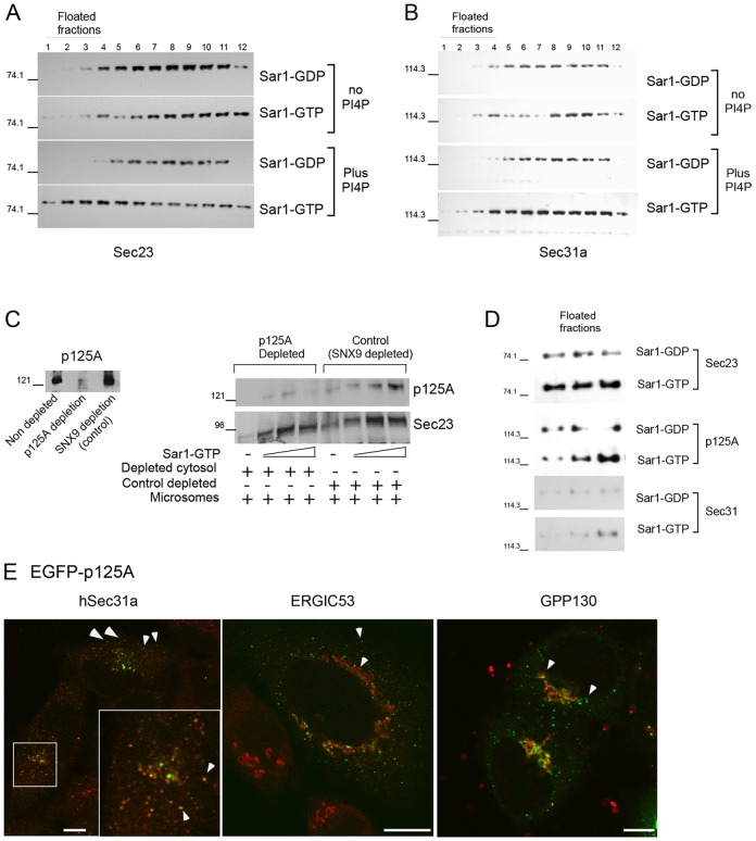
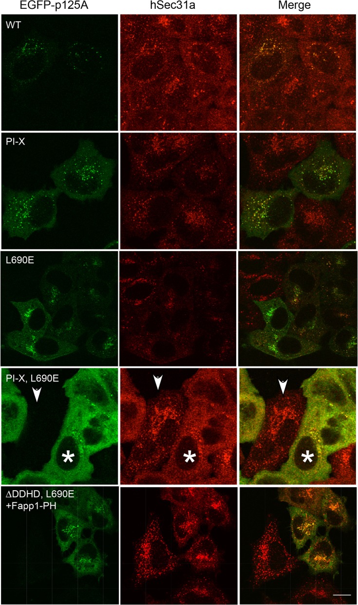
The inner and outer layers of COPII mediate cargo sorting and vesicle biogenesis. Sec16A and p125A (officially known as SEC23IP) proteins interact with both layers to control coat activity, yet the steps directing functional assembly at ER exit sites (ERES) remain undefined. By using temperature blocks, we find that Sec16A is spatially segregated from p125A-COPII-coated ERES prior to ER exit at a step that required p125A. p125A used lipid signals to control ERES assembly. Within p125A, we defined a C-terminal DDHD domain found in phospholipases and PI transfer proteins that recognized PA and phosphatidylinositol phosphates in vitro and was targeted to PI4P-rich membranes in cells. A conserved central SAM domain promoted self-assembly and selective lipid recognition by the DDHD domain. A basic cluster and a hydrophobic interface in the DDHD and SAM domains, respectively, were required for p125A-mediated functional ERES assembly. Lipid recognition by the SAM-DDHD module was used to stabilize membrane association and regulate the spatial segregation of COPII from Sec16A, nucleating the coat at ERES for ER exit.

Keywords: COPII; ERES; Phosphatidylinositol-4-phosphate; Sec23ip; p125A.


Figures

Fig. 1.
Fig. 1.
Sar1-dependent COPII and p125A recruitment requires PI4P. (A,B) Sar1-dependent COPII recruitment to floated liposomes is dependent on PI4P. Active (Sar1-GTP) or inactive (Sar1-GDP, both tested at 1 µg, 50 µl final volume) were incubated with rat liver cytosol and synthetic large unilamellar vesicles (LUV, 400 µM) composed of 45% phosphatidylcholine (PC), 35% phosphatidylethanolamine (PE), 10% phosphatidylserine (PS) and 10% cholesterol or 35% PC, 35% PE, 10% PS, 10% cholesterol and 10% PI4P at 26°C for 1 hour and floated onto a sucrose gradient (Bielli et al., 2005). Fractions were analyzed by western blot with antibodies against Sec23 (at 1:10,000 dilution) (A) or hSec31a (1:500) (B) (floated fractions as labeled). (C) Recruitment of Sec23 to ER membranes is not affected by depletion of p125A. p125A or SNX9 (control) was depleted from rat liver cytosol by immunoprecipitation and p125A depletion was verified using western blot, as indicated. Sar1-GTP-dependent Sec23 recruitment from control or p125A-depleted cytosol to ER microsomes was monitored. Sec23 was recruited by Sar1-GTP (50 ng, 500 ng and 1 µg, final volume 60 µL) in a dose-dependent manner in the absence (lanes 1–4) or presence (lanes 5–8) of p125A. (D) Floated PI4P-containing liposomes (fractions 1–3) from a COPII recruitment reaction (described in A) were probed for p125A (1:5000), Sec23 and Sec31. (E) Transiently expressed EGFP-tagged p125A colocalizes predominantly with hSec31a (as observed with the endogenous p125A protein) at ERES and was juxtaposed to ERGIC (ERGIC53) or cis-Golgi compartments (GPP130). Arrowheads indicate EGFP-p125A localizing with Sec31 (antibody dilution 1:100), adjacent to membranes containing ERGIC53 (1:100) and gpp130 (1:500). Scale bars: 5µm.
Fig. 2.
Fig. 2.
Cooperative lipid recognition by the SAM–DDHD module of p125A. (A) The DDHD domain targets PI4P-rich Golgi membrane in isolation. Transiently expressed EGFP–DDHD domain (779–989) dissociated from Sec31-containing ERES (arrowhead shows lack of colocalization between Sec31-stained ERES and localized EGFP–DDHD). The DDHD domain targets to the periphery of PI4P-enriched membranes and can be seen coating Golgi [right image, TGN46) (antibody dilution 1:500)] and ERGIC (ERGIC 53, middle image). Scale bars: 10 µm. (B-F) Selective lipid recognition is dependent on a module consisting of the DDHD and SAM domains. (B) 1 µg/ml of purified His-tagged p125A fragment (701–989) containing the DDHD domain, but not the SAM domain, was probed on lipid blot overlay using HRP-conjugated antibody against His6 (at 1:500 dilution). The fragment bound weakly to acidic lipids. (C) As in B: extending the fragment to contain the upstream SAM domain (643–989) conferred lipid selectivity to monophosphorylated PIs, PA, PS and PI(3,4)P2. (D) As in C: replacing a basic stretch of residues in the DDHD domain of the SAM–DDHD module with glutamic acid residues (850-KGRKR-854→EGEEE, termed PI-X) abolished lipid recognition. (E) The EGFP–DDHD domain is targeted to Golgi membranes (left), whereas EGFP–DDHDPI-X lost Golgi targeting (right). Scale bar: 5µm. (F) The EGFP-SAM domain transiently expressed in HeLa cells shows diffuse cytosolic distribution. Scale bar: 5µm. (G) The predicted structure of p125A SAM domain (643–704) (generated in Phyre) with red arrowhead indicating the position of a conserved leucine (690) within a hydrophobic dimer interface. (H) GST-SAM oligomerization is promoted by addition of Zn2+ and is sensitive to the L690E mutation. Buffer with or without the addition of 20 µM Zn(AOc)2 was added to GST-SAM or GST-SAML690E (10 µM of each), as indicated. Oligomerization was followed by the precipitation of the proteins from supernatant (S) to pellet (P) fractions (as indicated), using centrifugation and analysis on Coomassie-stained gels. (I) SAM and SAML690E domains (0.455 mM each) were incubated with 0.455 mM of Zn(AOc)2 and analyzed as in H.
Fig. 3.
Fig. 3.
p125A associated with COPII-coated ERES during temperature-induced traffic inhibition. (A) HeLa cells transiently expressing mRFP-p125A (8–12 hours) were maintained at 37°C, or incubated at 15°C or 10°C, as indicated, for 4 hours, then fixed and analyzed for localization with ERES (hSec31A). (B) Enlarged images of boxed areas in A, arrowheads indicate the extensive colocalization of p125A to ERES. (C) Colocalization of endogenous hSec31a and hSec24c (antibody at 1:100 dilution) in cells incubated at 10°C, as in A. Scale bars: 5µm.
Fig. 4.
Fig. 4.
ERES coated p125A segregates from Sec16A at low temperatures. (A) HeLa cells transiently expressing EGFP-Sec16A (8–12 hours) were maintained at 37°C, or incubated at 15°C or 10°C for 4 hours, and the localization of ERES (marked by hSec31a) and EGFP-Sec16A was determined. (B,C) Control HeLa cells (B) or cells transiently expressing mRFP-p125A (C) were incubated at 10°C and the localization of endogenous Sec16A (antibody at 1:1000 dilution), hSec31a and mRFP-p125A was determined, as indicated. Inset in C shows HeLa cells transiently expressing YFP-p58 analyzed for the localization of endogenous Sec16A and YFP-p58 during 10°C incubations. Arrowheads indicate hSec31 (A,B) or mRFP-p125A (C) sites, segregated from Sec16A. Scale bar: 5 µm.
Fig. 5.
Fig. 5.
PI4P binding by the SAM–DDHD module of p125A controls ERES assembly. The localization of transiently expressed (12–14 hours) full-length EGFP-p125A wild type, PI-X, L690E or the double mutant (p125AL690E,PI-X) and ERES (hSec31a) were analyzed in HeLa cells as indicated. The bottom panel shows the expression of a EGFP-p125A chimera where the DDHD domain has been substituted with the PH domain from Fapp1 in the backbone of an L690E mutant. EGFP-p125API-X or EGFP-p125AL690E mutants become partly cytosolic, whereas the double mutant p125API-X, L690E lost membrane localization and had completely disrupted ERES assembly. Membrane targeting and ERES assembly was partially restored in the Fapp1-PH-containing p125A chimera-expressing cells. Arrowheads indicate a non-transfected cell surrounded with cells expressing p125API-X, L690E (*). Scale bar: 10 µm.
Fig. 6.
Fig. 6.
Lipid recognition by p125A regulates Sec16A displacement from ERES. (A) HeLa cells transiently expressing (12–14 hours) mRFP-p125AL690E, ΔDDHD or mRFP-p125AL690E, ΔDDHD + Fapp1-PH were analyzed for hSec31 localization, as indicated. ERES [hSec31a (antibody dilution 1:200)] are disassembled in cells expressing mRFP p125AL690E, ΔDDHD (asterisks mark transfected cells), and mRFP-p125AL690E, ΔDDHD + Fapp1-PH and hSec31a are colocalized (arrows). (B) HeLa cells transiently expressing mRFP-p125A, mRFP-p125AL690E, ΔDDHD, EGFP-Sec16A, YFP-Sec23A, as indicated, for 24 hours were fixed and analyzed for the localization of transfected proteins. Note the segregation of EGFP-Sec16A from mRFP-p125A as opposed to the co-assembly of mRFP-p125A with YFP-Sec23a (arrow), and the collection of EGFP-Sec16A in mRFP-p125AL690E, ΔDDHD sites (arrows). Scale bar: 5 µm.
Fig. 7.
Fig. 7.
Functional SAM–DDHD module is required for steady-state ER-to-Golgi traffic. (A) Merge images showing the localization of endogenous p125A (green, antibody dilution 1:1000) and hSec31a (red) at ERES arrested at 10°C in control and p125A-depleted cells, as indicated (dsRNAi, supplementary material Table S2). Inset shows the localization of endogenous Sec16A (green) and hSec31a (red) under similar conditions. (B) Control or p125A-depleted cells were transfected with RNAi-resistant mRFP-p125Ar and traffic was arrested by incubation at 10°C. The localization of hSec31a was analyzed [merge images are shown, arrowhead indicates depleted cell next to rescued cells (asterisk)]. (C) HeLa cells stably expressing GFP-tagged N-acetylgalactosaminyltransferase-2 (GalNAcT2-GFP) were treated with control or p125A directed dsRNAi (supplementary material Table S2), as indicated, and visualized for GFP. Note the dramatic change in Golgi morphology (arrowheads) with loss of intact Golgi populations and the concomitant increase in shattered Golgi morphology. Scale bar: 5 µm. (D) Golgi morphology was quantified in HeLa cells by analyzing gpp130 localization. Typical observed Golgi morphologies are shown and color-coded, including intact (blue), dispersed (pink) and shattered (vesiculated, green). In some cells, Golgi morphology was not recognizable (labeled as missing, purple). (E) Analysis of p125A knockdown efficiency by western blots. Endogenous expression of p125A (middle panel, 1:2500) and actin (lower panel, 1:10,000) are shown. The upper panel shows the comparable expression of EGFP-p125A-resistant clones (EGFP-p125Ar; GFP antibody dilution 1:10,000) in control and KD cells. EGFP ran below the analyzed area and is not shown. The expression of EGFP-p125Ar (wild type) and EGFP-p125AL690E, PI-X were also detected by the p125A-specific antibody but required prolonged exposures due to partial transfection efficiency of KD cells. (F) The fractional distribution of Golgi morphologies in control, p125A-depleted and rescued cell populations with EGFP-p125Ar and EGFP-p125API-X, L690Er, as indicated. The percentage of cells with either intact Golgi (blue), dispersed Golgi (pink), shattered Golgi (green) or missing Golgi (purple) under each treatment condition is shown. (G) Statistical analysis of intact Golgi morphology in control and KD cells (mean±s.d.). Three experiments were performed for each condition. Ten images were collected from each experiment and Golgi phenotypes were determined for all cells expressing EGFP-tagged proteins (93–220 cells per group). Unpaired Student's t-test was used to test for significant differences between groups.
Fig. 8.
Fig. 8.
The COPII budding cascade at ERES. (A–C) Following initial association of COPII layers with Sec16 on ER membranes (A), the binding of PI4P by p125A promotes the displacement of Sec16 from COPII inner and outer layers to allow for effective linking between coat layers. This drives coat retention (B) while enhancing GTP hydrolysis to support later steps, including Sar1 exchange by Bet3 on Sec23, and Sar1-induced vesicle neck constriction and fission (C).

Similar articles

Cited by

References

    1. Antonny B., Schekman R. (2001). ER export: public transportation by the COPII coach. Curr. Opin. Cell Biol. 13, 438–443 10.1016/S0955-0674(00)00234-9 - DOI - PubMed
    1. Antonny B., Madden D., Hamamoto S., Orci L., Schekman R. (2001). Dynamics of the COPII coat with GTP and stable analogues. Nat. Cell Biol. 3, 531–537 10.1038/35078500 - DOI - PubMed
    1. Aridor M., Bannykh S. I., Rowe T., Balch W. E. (1995). Sequential coupling between COPII and COPI vesicle coats in endoplasmic reticulum to Golgi transport. J. Cell Biol. 131, 875–893 10.1083/jcb.131.4.875 - DOI - PMC - PubMed
    1. Aridor M., Weissman J., Bannykh S., Nuoffer C., Balch W. E. (1998). Cargo selection by the COPII budding machinery during export from the ER. J. Cell Biol. 141, 61–70 10.1083/jcb.141.1.61 - DOI - PMC - PubMed
    1. Aridor M., Fish K. N., Bannykh S., Weissman J., Roberts T. H., Lippincott-Schwartz J., Balch W. E. (2001). The Sar1 GTPase coordinates biosynthetic cargo selection with endoplasmic reticulum export site assembly. J. Cell Biol. 152, 213–230 10.1083/jcb.152.1.213 - DOI - PMC - PubMed

Publication types

MeSH terms