. 2014 Apr 15;127(Pt 8):1779-91.
doi: 10.1242/jcs.139014. Epub 2014 Feb 12.

α-catenin cytomechanics--role in cadherin-dependent adhesion and mechanotransduction

Affiliations

α-catenin cytomechanics--role in cadherin-dependent adhesion and mechanotransduction

Adrienne K Barry et al. J Cell Sci. .

Abstract

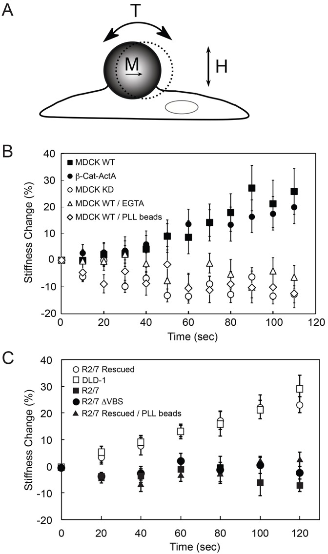
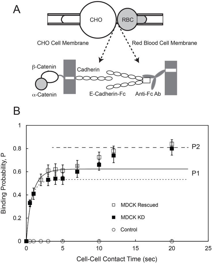
The findings presented here demonstrate the role of α-catenin in cadherin-based adhesion and mechanotransduction in different mechanical contexts. Bead-twisting measurements in conjunction with imaging, and the use of different cell lines and α-catenin mutants reveal that the acute local mechanical manipulation of cadherin bonds triggers vinculin and actin recruitment to cadherin adhesions in an actin- and α-catenin-dependent manner. The modest effect of α-catenin on the two-dimensional binding affinities of cell surface cadherins further suggests that force-activated adhesion strengthening is due to enhanced cadherin-cytoskeletal interactions rather than to α-catenin-dependent affinity modulation. Complementary investigations of cadherin-based rigidity sensing also suggest that, although α-catenin alters traction force generation, it is not the sole regulator of cell contractility on compliant cadherin-coated substrata.

Keywords: Adhesion; Cadherin; Mechanotransduction; α-Catenin.


Figures

Fig. 1.
Fig. 1.
Western blots of α-catenin expression in MDCK and DLD-1 cell lines. Whole-cell lysates from MDCK WT (parental), MDCK KD (clone number 1) and MDCK Rescued (clone number 10) cells (left) and DLD-1 (parental), R2/7 and R2/7 Rescued cells (right) were separated by SDS-PAGE and blotted for α-catenin, GAPDH and tubulin.
Fig. 2.
Fig. 2.
α-Catenin is required for acute cadherin-dependent mechanotransduction. (A) Schematic of the magnetic twisting cytometry experiment. Ligand-coated ferromagnetic beads are magnetized with a magnetic moment (M) parallel to the substrate and subjected to an oscillating field (H). The orthogonal applied field generates a torque (T) on the bead, causing a bead displacement. (B) MTC measurements of force-induced cell stiffening were performed using canine E-cad-Fc-coated beads to probe MDCK WT cells in the absence (black squares) and presence of 4 mM EGTA (white triangles). In controls, MDCK WT cells were probed with PLL-coated beads (white diamonds). E-cad-Fc-coated beads also probed MDCK WT cells in which α-catenin has been stably knocked down (MDCK KD, white circles), and MDCK KD cells expressing β-cat-ActA (black circles). (C) MTC measurements using E-cad-Fc-coated beads to probe DLD-1 cells (white squares), α-catenin-null cells (R2/7, black squares), R2/7 cells rescued with mouse GFP–α-catenin (R2/7 Rescued, white circles) and R2/7 cells rescued with mouse GFP–α-catenin lacking the vinculin-binding site (R2/7 ΔVBS, black circles). In controls, R2/7 Rescued cells were probed with PLL-coated beads (black triangles). Each time point represents the mean±s.d. of >200 beads (one bead per cell).
Fig. 3.
Fig. 3.
Kinetics of E-cadherin-mediated binding between an MDCK cell and an RBC modified with E-cad-Fc. (A) Schematic of the micropipette aspiration experiment. A cell expressing full-length cadherin is aspirated into a pipette (left) and repetitively brought into contact with an RBC modified with Fc-tagged canine E-cadherin extracellular domains (E-cad-Fc), which are captured and oriented by anti-Fc antibody covalently bound to the RBC (right). (B) The time-dependent binding probability (P) versus cell–cell contact time measured between RBCs modified with E-cad-Fc and MDCK KD cells (black squares) or MDCK Rescued cells (white squares). The solid line is the nonlinear least squares fit of Eqn 1 to data for the first binding step obtained with MDCK Rescued cells, with best-fit parameters given in the text. The dotted line is the fit to data obtained with MDCK KD cells, with best-fit parameters given in the text. The dashed line indicates the limiting binding probability P2 determined with both MDCK Rescued and MDCK KD cells. Control data (white circles) were measured between MDCK Rescued cells and RBCs modified with anti-human IgG (Fc) antibody without bound E-cad-Fc.
Fig. 4.
Fig. 4.
Force-dependent distributions of α-catenin, F-actin and vinculin imaged at cell–cell and bead–cell junctions. (A) Immunostained R2/7 Rescued, R/27 and R2/7 ΔVBS cells before applying shear stress through E-cad-Fc-coated beads. R2/7 Rescued and R2/7 ΔVBS cells overexpress GFP–α-catenin and GFP–α-catenin-ΔVBS, respectively. R2/7 cells are deficient in α-catenin. Representative images reveal α-catenin (green) and F-actin (orange) at cell–cell junctions at the basal plane. Scale bars: 10 µm. (B) α-Catenin and F-actin distributions at PLL-coated beads bound to the apical surface of R2/7 Rescued cells, before or after bond shear. Images of individual beads were cropped and enlarged from the boxed regions of original images (shown in supplementary material Fig. S2A). The first DIC image (top left) indicates the region of interest (yellow circles) used to quantify changes in protein distributions at bead–cell junctions. Images are representative of >35 different bead–cell pairs. (C) R2/7 Rescued cells stained for F-actin, before or after cadherin bond shear. (D) R2/7 Rescued cells stained for vinculin. (E,F) R/27 cells stained for F-actin or vinculin, respectively. (G,H) R2/7 ΔVBS cells stained for F-actin or vinculin, respectively. Original images for C–H are shown in supplementary material Fig. S2B. Images are representative of >35 different bead–cell pairs. Scale bars: 5 µm.
Fig. 5.
Fig. 5.
E-cadherin distributions at cell–cell and bead–cell junctions in R2/7 cells expressing GFP–α-catenin or GFP–α-catenin-ΔVBS. (A) R2/7 Rescued and R2/7 ΔVBS cells immunostained for E-cadherin, before or after applying cadherin bond shear. Cell–cell junctions were imaged at the basal plane of cell monolayers for E-cadherin. Representative DIC images show beads attached to cells, and the corresponding fluorescence images show α-catenin (green) and E-cadherin (orange). Scale bars: 10 µm. (B) Mean fluorescence intensity of α-catenin and E-cadherin at E-cadherin bead–cell junctions in R2/7 Rescued (left; α-catenin, n≥208; E-cadherin, n≥55) and R2/7 ΔVBS cells (right; α-catenin, n≥44; E-cadherin, n≥44).
Fig. 6.
Fig. 6.
Shear-induced changes in α-catenin, vinculin and F-actin at E-cadherin bead–cell junctions. (A) Change in mean fluorescence intensity, relative to non-loading conditions, of proteins within rings extending 1.0–1.5 µm from the bead edge. A mask designating a filled outline of the bead was defined, based on DIC images. Data represent R2/7 Rescued (α-catenin, n≥208; vinculin, n≥85; F-actin, n≥60), R/27 (vinculin, n≥55; F-actin, n≥35) and R2/7 ΔVBS (α-catenin, n≥86; vinculin, n≥42; F-actin, n≥44) cells bound to E-cad-Fc beads. The control was obtained with PLL beads bound to R2/7 Rescued cells (α-catenin, n≥119; vinculin, n≥58; F-actin, n≥61). (B) Fluorescence intensities of proteins before and after applied load in R2/7 Rescued cells bound to E-cad-Fc beads. Left: F-actin, ***P = 2×10⁻⁸, n≥60; α-catenin, P = 0.64, n≥208. Right: vinculin, ***P = 2×10⁻⁵, n≥85; α-catenin, P = 0.64, n≥208. (C) Intensity levels at E-cad-Fc beads on R2/7 ΔVBS cells, with and without load. Left: F-actin, n≥44; α-catenin, n≥44. Right: vinculin, n≥42; α-catenin, n≥42. (D) Intensity levels at PLL beads on R2/7 Rescued cells. Left: F-actin, n≥61; α-catenin, n≥61. Right: vinculin, n≥58; α-catenin, n≥58. All error bars represent s.e.m.
Fig. 7.
Fig. 7.
α-Catenin modulates cadherin-mediated traction forces. (A) Root mean square (RMS) traction forces (Pa) exerted by MDCK KD and MDCK Rescued cells on soft (1 kPa) and rigid (34 kPa) hydrogels with covalently immobilized and oriented E-cad-Fc. For each condition, n≥4; *P<0.05, **P<0.01, ***P<0.001. Error bars represent s.e.m. (B) Representative traction force maps of MDCK KD and MDCK Rescued cells on soft and rigid hydrogels. (C) RMS traction forces exerted by R2/7, R2/7 ΔVBS, R2/7 Rescued and DLD-1 cells on soft (1 kPa), semi-rigid (9 kPa) and rigid (34 kPa) hydrogels with covalently bound E-cad-Fc. All measurements were done in the presence of integrin-blocking antibodies. For each condition, n≥10. Representative traction force maps are in supplementary material Fig. S4. (D) RMS traction forces exerted by MDCK Rescued and MDCK KD cells on 34-kPa gels coated with E-cad-Fc, after treatment with blebbistatin or cytochalasin D (Cyto D). For each condition, n≥5; *P<0.05, **P<0.01, ***P<0.001. All error bars represent s.e.m. (E) Immunofluorescence images of paxillin and F-actin at the basal plane of R2/7 and R2/7 Rescued cells on 34 kPa gels modified with E-cad-Fc. Scale bars: 10 µm.

References

    1. Bajpai S., Correia J., Feng Y., Figueiredo J., Sun S. X., Longmore G. D., Suriano G., Wirtz D. (2008). alpha-Catenin mediates initial E-cadherin-dependent cell-cell recognition and subsequent bond strengthening. Proc. Natl. Acad. Sci. USA 105, 18331–18336 10.1073/pnas.0806783105 - DOI - PMC - PubMed
    1. Beningo K. A., Wang Y. L. (2002). Flexible substrata for the detection of cellular traction forces. Trends Cell Biol. 12, 79–84 10.1016/S0962-8924(01)02205-X - DOI - PubMed
    1. Beningo K. A., Lo C. M., Wang Y. L. (2002). Flexible polyacrylamide substrata for the analysis of mechanical interactions at cell-substratum adhesions. Methods Cell Biol. 69, 325–339 10.1016/S0091-679X(02)69021-1 - DOI - PubMed
    1. Benjamin J. M., Kwiatkowski A. V., Yang C., Korobova F., Pokutta S., Svitkina T., Weis W. I., Nelson W. J. (2010). AlphaE-catenin regulates actin dynamics independently of cadherin-mediated cell-cell adhesion. J. Cell Biol. 189, 339–352 10.1083/jcb.200910041 - DOI - PMC - PubMed
    1. Braga V. M., Del Maschio A., Machesky L., Dejana E. (1999). Regulation of cadherin function by Rho and Rac: modulation by junction maturation and cellular context. Mol. Biol. Cell 10, 9–22 10.1091/mbc.10.1.9 - DOI - PMC - PubMed

Publication types

LinkOut - more resources