. 2014 Apr 15;127(Pt 8):1840-53.
doi: 10.1242/jcs.144550. Epub 2014 Feb 12.

Non-muscle Mlck is required for β-catenin- and FoxO1-dependent downregulation of Cldn5 in IL-1β-mediated barrier dysfunction in brain endothelial cells

Affiliations

Non-muscle Mlck is required for β-catenin- and FoxO1-dependent downregulation of Cldn5 in IL-1β-mediated barrier dysfunction in brain endothelial cells

Richard S Beard Jr et al. J Cell Sci. .

Abstract

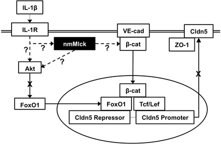
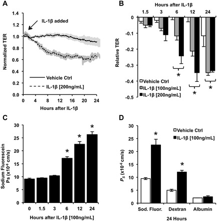
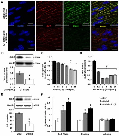
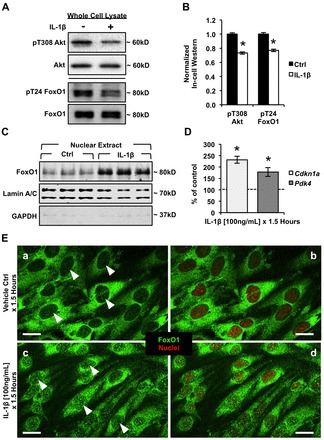
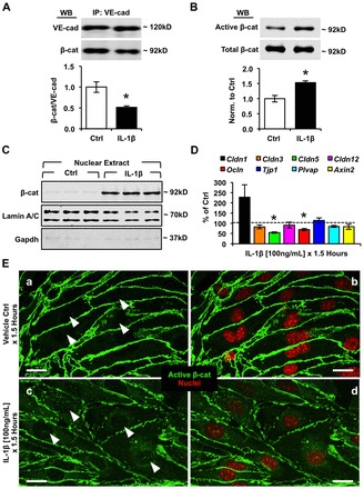
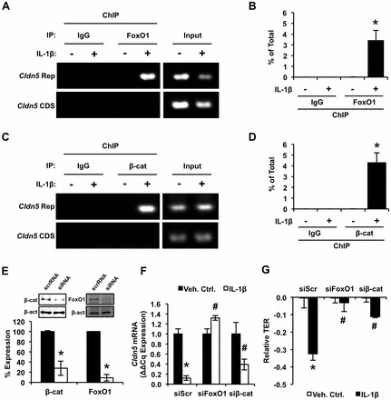
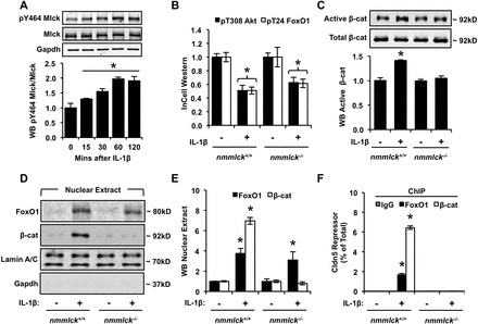
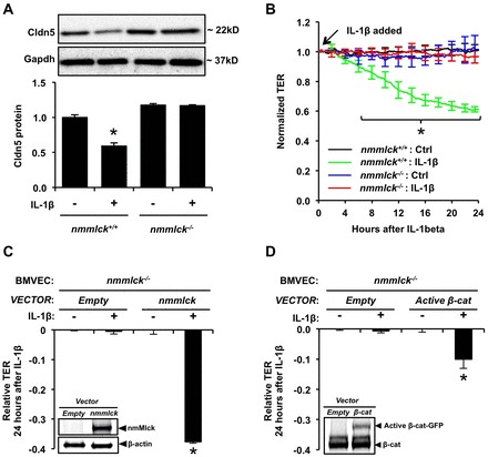
Aberrant elevation in the levels of the pro-inflammatory cytokine interleukin-1β (IL-1β) contributes to neuroinflammatory diseases. Blood-brain barrier (BBB) dysfunction is a hallmark phenotype of neuroinflammation. It is known that IL-1β directly induces BBB hyperpermeability but the mechanisms remain unclear. Claudin-5 (Cldn5) is a tight junction protein found at endothelial cell-cell contacts that are crucial for maintaining brain microvascular endothelial cell (BMVEC) integrity. Transcriptional regulation of Cldn5 has been attributed to the transcription factors β-catenin and forkhead box protein O1 (FoxO1), and the signaling molecules regulating their nuclear translocation. Non-muscle myosin light chain kinase (nmMlck, encoded by the Mylk gene) is a key regulator involved in endothelial hyperpermeability, and IL-1β has been shown to mediate nmMlck-dependent barrier dysfunction in epithelia. Considering these factors, we tested the hypothesis that nmMlck modulates IL-1β-mediated downregulation of Cldn5 in BMVECs in a manner that depends on transcriptional repression mediated by β-catenin and FoxO1. We found that treating BMVECs with IL-1β induced barrier dysfunction concomitantly with the nuclear translocation of β-catenin and FoxO1 and the repression of Cldn5. Most importantly, using primary BMVECs isolated from mice null for nmMlck, we identified that Cldn5 repression caused by β-catenin and FoxO1 in IL-1β-mediated barrier dysfunction was dependent on nmMlck.

Keywords: Blood–brain barrier; Claudin-5; FoxO1; IL-1β; Neuroinflammation; Non-muscle myosin light chain kinase; β-catenin.


Figures

Fig. 1.
Fig. 1.
Diagram of signaling pathways through which non-muscle myosin light chain kinase (nmMlck) might be involved in IL-1β-mediated BMVEC barrier dysfunction. We hypothesize that nmMlck modulates IL-1β-mediated downregulation of Cldn5 and dysfunction of the BMVEC barrier in a manner that promotes the nuclear translocation of β-catenin and the repression of the Cldn5 gene. Question marks and dashed lines indicate unknown signaling mechanisms, whereas solid lines indicate previously established mechanisms.
Fig. 2.
Fig. 2.
Treating brain microvascular endothelial cells (bEnd.3 cells) with IL-1β induces a time- and dose-dependent decrease in TER while increasing size-selective monolayer permeability. (A) ECIS tracings of monolayers treated with vehicle control (0.1% BSA in PBS) or IL-1β (200 ng/ml). Tracings indicate the mean resistance normalized to t = 0 (indicated by arrow) and shading indicates s.e.m. (B) Statistical analysis of relative TER decline at the indicated time points during the treatment of bEnd.3 monolayers with IL-1β (either 100 or 200 ng/ml). (C) Time-course of the IL-1β-mediated increase in monolayer permeability to sodium fluorescein. (D) Monolayers were treated with vehicle control or IL-1β (100 ng/ml) for 24 hours, then transwell permeability assays were performed to determine permeability coefficients (PS) for the following molecules: sodium fluorescein (0.376 kDa; Stokes' radius≈0.45 nm), TRITC–dextran (4.4 kDa; Stokes' radius≈1.4 nm) and FITC–albumin (66.4 kDa; Stokes' radius≈3.48 nm). Data are presented as the mean±s.e.m. *P<0.05 compared with vehicle control or t = 0; one-way ANOVA with Dunnett's post hoc analysis.
Fig. 3.
Fig. 3.
IL-1β decreases Cldn5 expression in bEnd.3 cells in a manner consistent with IL-1β-mediated barrier dysfunction. (A) Confocal immunofluorescence analysis of the tight junction proteins Cldn5 and zonula occludens-1 (ZO-1) after a 24-hour treatment with IL-1β (100 ng/ml), compared with vehicle control. Scale bars: 20 µm. Cldn5 and ZO-1 are colocalized at cell–cell contacts in vehicle-control-treated cells. In IL-1β-treated cells, no obvious expression or localization changes were observed for ZO-1; however, IL-1β induced a significant reduction in Cldn5 expression at cell–cell contacts. (B) Western blotting for Cldn5 in cell lysates demonstrated that treatment with IL-1β (100 ng/ml) induced ∼2-fold decrease in total Cldn5 protein expression. (C) ICWs were used to determine the time-course for the IL-1β-mediated decrease in Cldn5 protein expression. (D) Real-time PCR results demonstrate that IL-1β induced downregulation of Cldn5 mRNA that preceded the IL-1β-mediated decrease in Cldn5 protein expression. (E) Western blotting providing confirmation and quantification of Cldn5 knockdown that was obtained by treatment with targeted siRNA (siCldn5) or scrambled siRNA (siScr). (F) Permeability coefficients (PS) for sodium fluorescein, TRITC-dextran and FITC-albumin were measured in Cldn5-deficient BMVEC monolayers (siCldn5) with or without IL-1β (100 ng/ml) treatment for 24 hours, and were compared with those of siScr monolayers. Similar to IL-1β treatment, Cldn5 deficiency increased the permeability of the monolayer to sodium fluorescein and TRITC–dextran, but not to FITC–albumin. Moreover, treating Cldn5-deficient monolayers with IL-1β further increased the permeability of the monolayer to sodium fluorescein and TRITC–dextran, whereas FITC–albumin permeability remained unchanged. Data are presented as the mean±s.e.m. *P<0.05 compared with the vehicle control, the t = 0 time-point or siScr; #P<0.05 compared with siCldn5 alone; Student's t-test or one-way ANOVA with Dunnett's (C,D) and Tukey's (F) post hoc analysis.
Fig. 4.
Fig. 4.
IL-1β treatment is associated with Akt inactivation, resulting in FoxO1 nuclear accumulation and FoxO1-dependent regulation of gene expression. (A) Representative western blots demonstrate that IL-1β (100 ng/ml for 1.5 hours) inactivated Akt, as indicated by a decrease in phosphorylation of T308. Consistent with inactivation of Akt, IL-1β treatment also led to decreased phosphorylation of FoxO1 on T24. (B) ICWs were used to quantify IL-1β-mediated dephosphorylation of Akt and FoxO1. (C) Western blot analysis of nuclear lysates demonstrated a significant increase in the nuclear accumulation of FoxO1 in response to IL-1β treatment. Lamin A/C was used as a loading control and Gapdh was used to determine the efficacy of nuclear fractionation. (D) Real-time PCR results showed upregulation of two known FoxO1 transcriptional targets, Cdkn1a and Pdk4. Data are presented as the mean±s.e.m. *P<0.05 compared with vehicle control; Student's t-test. (E) Confocal immunofluorescence analysis revealed an increased nuclear accumulation of FoxO1 in response to IL-1β treatment (c,d) compared with cells treated with vehicle control (a,b). Arrowheads represent the location of nuclei in images without nuclear staining. Scale bars: 20 µm.
Fig. 5.
Fig. 5.
Treatment of bEnd.3 cells with IL-1β leads to uncoupling of the VE-cadherin–β-catenin complex, an increase in the pool of active β-catenin, and nuclear localization of β-catenin. (A) Co-immunoprecipitation experiments demonstrated that there was a decrease in the amount of β-catenin (β-cat) that precipitated with VE-cadherin (VE-cad) after IL-1β treatment (100 ng/ml, 1.5 hours). (B) Western blotting for active β-catenin (clone 8E7) demonstrated an increase in active β-catenin immunolabeling after IL-1β treatment. Data represent the mean±s.e.m. (C) Western blot analysis of the nuclear fraction showed a significant increase in nuclear accumulation of β-catenin in response to IL-1β treatment. Lamin A/C was used as a loading control, and Gapdh was used to determine the efficacy of nuclear fractionation. The control blots showing Lamin A/C and Gapdh are duplicates of the controls shown in Fig. 4C, as these experiments were performed on the same nuclear extracts and western blots. (D) mRNA levels were determined for Cldn1, Cldn3, Cldn5, Cldn12, Ocln, Tjp1, Plvap and Axin2 from bEnd.3 cells that were treated with IL-1β. Data represent mean mRNA levels (±s.e.m.), shown as a percentage of vehicle control. *P<0.05 compared with vehicle control; Student's t-test. (E) Confocal immunocytochemistry demonstrates increased nuclear accumulation of β-catenin in response to IL-1β treatment (c without nuclei staining, d with nuclei staining) compared with vehicle control (a without nuclei staining, b with nuclei staining). Arrowheads represent the location of nuclei in images without nuclei staining. Scale bars: 20 µm.
Fig. 6.
Fig. 6.
IL-1β requires the recruitment of FoxO1 and β-catenin to the Cldn5 repressor to reduce Cldn5 expression. 1.5 hours after treating bEnd.3 cells with IL-1β (100 ng/ml) or vehicle control, bound chromatin was immunoprecipitated (ChIP) with anti-FoxO1, anti-β-catenin or IgG isotype-control antibodies. ChIP samples were assayed by using PCR with primers for the putative Cldn5 repressor region, whereas primers for the Cldn5 coding sequence (CDS) were used as negative controls for non-specific DNA precipitation. (A,C) Qualitative PCR results from ChIP assays. (B,D) Quantitative RT-PCR results from ChIP assays. ΔCq (or percentage of total) for the Cldn5 repressor was calculated by normalizing to input. (E) Western blotting confirmation and quantification of gene knockdown using targeted siRNA against β-catenin (siβ-cat), FoxO1 (siFoxO1) or scrambled control (siScr). FoxO1 and β-catenin knockdown attenuated IL-1β-induced downregulation of Cldn5 mRNA at 1.5 hours after treatment (F) and barrier dysfunction at 24 hours (G). Data represent the mean±s.e.m. *P<0.05 compared with control, #P<0.05 compared with siScr treated with IL-1β; Student's t-test.
Fig. 7.
Fig. 7.
nmMlck is required for β-catenin and FoxO1 occupancy of the Cldn5 repressor. Primary BMVECs were isolated from cortices of wild-type (nmmlck+/+) and nmMlck homozygous knockout (nmmlck−/−) mice. (A) Treating BMVECs with IL-1β (100 ng/ml) acutely (within 15 minutes) activated nmMlck, as indicated by increased phosphorylation of Y464 in a time-dependent manner. (B) ICWs were used to assess IL-1β-mediated inactivation of the Akt–FoxO1 pathway in BMVECs, by using phosphospecific antibodies against T308 on Akt (pT308 Akt) and T24 on FoxO1 (pT24 FoxO1). Antibodies against total Akt and FoxO1 were used for co-labeling, and the measurements were used to normalize phosphospecific signals. (C) IL-1β (100 ng/ml, 1.5 hours) increased the ratio of active β-catenin (hypophosphorylated at GSK3β sites Ser37 and Thr41) to total β-catenin in BMVECs isolated from nmmlck+/+ mice, but not in those isolated from nmmlck−/− mice. *P<0.05 compared with control. (D,E) Consistent with the inactivation and activation signals on Akt, FoxO1 and β-catenin, nmMlck deficiency attenuated IL-1β-mediated nuclear accumulation of β-catenin, but not nuclear accumulation of FoxO1. (F) Although IL-1β treatment led to nuclear accumulation of FoxO1 in both nmmlck+/+ and nmmlck−/− BMVECs, the absence of β-catenin in the nuclei of nmmlck−/− BMVECs treated with IL-1β prevented the binding of FoxO1 to the Cldn5 repressor. Data represent the mean±s.e.m. *P<0.05 compared with all other groups; Student's t-test.
Fig. 8.
Fig. 8.
nmMlck deficiency attenuates IL-1β-mediated dysfunction of the brain endothelial barrier and Cldn5 downregulation. (A) Western blotting for Cldn5 in lysates from BMVECs treated with IL-1β (100 ng/ml, 24 hours) revealed that IL-1β-mediated Cldn5 downregulation is dependent on nmMlck. (B) ECIS experiments demonstrated that BMVECs from nmmlck+/+ mice respond to IL-1β in a similar fashion to bEnd.3 cells. (C) Ectopic expression of nmMlck in nmmlck−/− BMVECs reverts the attenuation of IL-1β-mediated barrier dysfunction. (D) Consistent with the evidence that activation of β-catenin is dependent on nmMlck, ectopic expression of active β-catenin in nmmlck−/− BMVECs also restores IL-1β-mediated barrier dysfunction. Data represent the mean±s.e.m. *P<0.05 compared with all other groups; Student's t-test.

Similar articles

Cited by

References

    1. Banks W. A. (1999). Physiology and pathology of the blood–brain barrier: implications for microbial pathogenesis, drug delivery and neurodegenerative disorders. J. Neurovirol. 5, 538–555 10.3109/13550289909021284 - DOI - PubMed
    1. Barbieri S. S., Weksler B. B. (2007). Tobacco smoke cooperates with interleukin-1beta to alter beta-catenin trafficking in vascular endothelium resulting in increased permeability and induction of cyclooxygenase-2 expression in vitro and in vivo. FASEB J. 21, 1831–1843 10.1096/fj.06-7557com - DOI - PubMed
    1. Barbieri S. S., Ruggiero L., Tremoli E., Weksler B. B. (2008). Suppressing PTEN activity by tobacco smoke plus interleukin-1beta modulates dissociation of VE-cadherin/beta-catenin complexes in endothelium. Arterioscler. Thromb. Vasc. Biol. 28, 732–738 10.1161/ATVBAHA.107.159434 - DOI - PubMed
    1. Basu A., Krady J. K., Levison S. W. (2004). Interleukin-1: a master regulator of neuroinflammation. J. Neurosci. Res. 78, 151–156 10.1002/jnr.20266 - DOI - PubMed
    1. Bates D. O. (2010). Vascular endothelial growth factors and vascular permeability. Cardiovasc. Res. 87, 262–271 10.1093/cvr/cvq105 - DOI - PMC - PubMed

Publication types

MeSH terms