Comparative Study
. 1988 May;50(5):1447-55.
doi: 10.1111/j.1471-4159.1988.tb03029.x.

Protein tyrosine kinase activity and its endogenous substrates in rat brain: a subcellular and regional survey

Affiliations
Comparative Study

Protein tyrosine kinase activity and its endogenous substrates in rat brain: a subcellular and regional survey

A A Hirano et al. J Neurochem. 1988 May.

Abstract

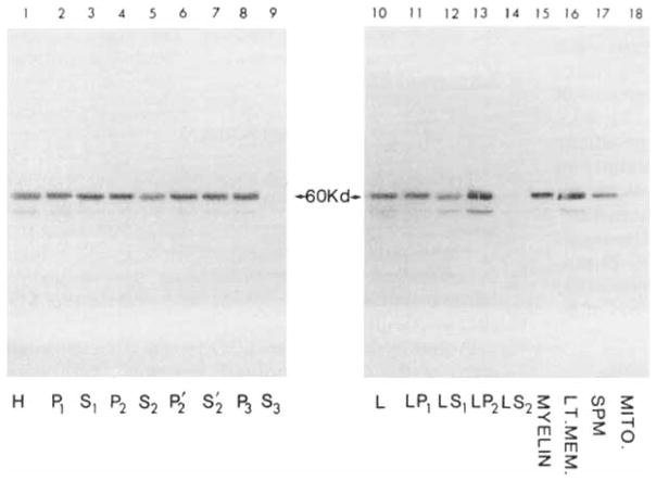
The rat CNS contains high levels of tyrosine-specific protein kinases that specifically phosphorylate the tyrosine-containing synthetic peptide poly(Glu80,Tyr20). The phosphorylation of this peptide is rapid and occurs with normal Michaelis-Menten kinetics. Using this peptide to assay for enzyme activity, we have measured the protein tyrosine kinase activity in homogenates from various regions of rat CNS. A marked regional distribution pattern was observed, with high activity present in cerebellum, hippocampus, olfactory bulb, and pyriform cortex, and low activity in the pons/medulla and spinal cord. The distribution of protein tyrosine kinase activity was examined in various subcellular fractions of rat forebrain. The majority of the activity was associated with the particulate fractions, with enrichment in the crude microsomal (P3) and crude synaptic vesicle (LP2) fractions. Moreover, the subcellular distribution of pp60csrc, a well-characterized protein tyrosine kinase, was examined by immunoblot analysis using an affinity-purified antibody specific for pp60csrc. The subcellular distribution of pp60csrc paralleled the overall protein tyrosine kinase activity. In addition, using an antibody specific for phosphotyrosine, endogenous substrates for protein tyrosine kinases were demonstrated on immunoblots of homogenates from the various regions and the subcellular fractions. The immunoblots revealed numerous phosphotyrosine-containing proteins that were present in many of the CNS regions examined and were associated with specific subcellular fractions. The differences in tyrosine-specific protein kinase activity, and in phosphotyrosine-containing proteins, observed in various regional areas and subcellular fractions may reflect specific functional roles for protein tyrosine kinase activity in mammalian brain.


Figures

FIG. 1
FIG. 1
Demonstration of tyrosine-specific protein kinase activity in rat forebrain. The phosphorylation reaction was carried out under standard conditions for 30 min with 50 μg of protein in the absence (−) or presence (+) of 0.5 mg/ml poly(Glu80, Tyr20). Left: SDS-PAGE analysis. Right: Phosphoamino acid analysis. The spots near the origin of the TLC plates are unhydrolyzed phosphopeptides.
FIG. 2
FIG. 2
Lineweaver–Burk plots of the phosphorylation of poly(Glu80,Tyr20) by the endogenous protein tyrosine kinase activity of rat forebrain. Initial rates were measured under standard conditions for 5 min, using the filter paper assay. ATP concentration (A) and Poly(Glu80,Tyr20) concentration (B) were varied as indicated.
FIG. 3
FIG. 3
Regional distribution of protein tyrosine kinase activity in rat CNS. The material dissected from the various regions was homogenized and the protein tyrosine kinase activity was measured under standard conditions, using the filter paper assay. These data are the averages from three different dissections. The results represent picomoles of phosphate incorporated into the synthetic substrate, presented as means ± SEM. Olf. bulb, olfactory bulb; Olf. tube., olfactory tubercle; Caud-put., caudatoputamen; N. acc., nucleus accumbens; Glob, pall., globus pallidus; Hypothal., hypothalamus; Thal., thalamus; Septum, septum; Hippo., hippocampus; Pyr. ctx., pyriform cortex; Neoctx., neocortex; V. Mes., ventral mesencephalon; M. Mes., middle mesencephalon; D. Mes., dorsal mesencephalon; Cbllm., cerebellum; Pons/med., pons/medulla; Sp. cord, spinal cord.
FIG. 4
FIG. 4
Subcellular distribution of protein tyrosine kinase activity in rat forebrain. Protein tyrosine kinase activity was assayed under standard conditions, using the filter paper assay. These data are the averages of three separate subcellular fractionations, assayed in duplicate. Results represent picomoles of phosphate incorporated into the synthetic substrate, presented as means ± SEM. The various fractions are as defined in Materials and Methods.
FIG. 5
FIG. 5
Subcellular distribution of pp60csrc in rat forebrain. Each subcellular fraction (50 μg of protein) was subjected to SDS-PAGE on 8% gels, and transferred onto nitrocellulose. The nitrocellulose filters were probed with the anti-pp60csrc antibodies and developed with alkaline phosphatase-conjugated goat anti-rabbit IgG. The various fractions are as defined in Materials and Methods.
FIG. 6
FIG. 6
Regional distribution of phosphotyrosine-containing proteins in rat CNS. An aliquot (50 μg of protein) of the homogenate from each region was phosphorylated under standard conditions using nonradioactive ATP, subjected to SDS-PAGE on 8% gels, and transferred onto nitrocellulose. The nitrocellulose filters were probed with the anti-phosphotyrosine antibodies, developed with 125I-Protein A, and subjected to autoradiography. For abbreviations used see legend to Fig. 3.
FIG. 7
FIG. 7
Subcellular distribution of phosphotyrosine-containing proteins in rat forebrain. Each subcellular fraction (50 μg of protein) was phosphorylated under standard conditions using nonradioactive ATP, subjected to SDS-PAGE on 8% gels, and transferred onto nitrocellulose. The nitrocellulose filters were probed with the anti-phosphotyrosine antibodies, developed with 125I-Protein A, and subjected to autoradiography.

References

    1. Albuquerque EX, Deshpande SS, Aracava Y, Alkondon M, Daly JW. A possible involvement of cyclic AMP in the expression of desensitization of the nicotinic acetylcholine receptor. A study with forskolin and its analogs. FEBS Lett. 1986;199:113–120. - PubMed
    1. Barnekow A, Schartl M, Anders F, Bauer H. Identification of a fish protein associated with a kinase activity and related to the Rous sarcoma virus transforming protein. Cancer Res. 1982;42:2429–2433. - PubMed
    1. Braun S, Raymond WE, Racker E. Synthetic tyrosine polymers as substrates and inhibitors of tyrosine-specific protein kinases. J Biol Chem. 1984;259:2051–2054. - PubMed
    1. Braun S, Ghany MA, Lettieri JA, Racker E. Partial purification and characterization of protein tyrosine kinases from normal tissues. Arch Biochem Biophys. 1986;247:424–432. - PubMed
    1. Browning MD, Huganir R, Greengard P. Protein phosphorylation and neuronal function. J Neurochem. 1985;45:11–23. - PubMed

Publication types

MeSH terms

Substances

LinkOut - more resources