. 2014 May;58(2-3):224-33.
doi: 10.1007/s12026-014-8491-6.

Effects of serum and plasma matrices on multiplex immunoassays

Affiliations

Effects of serum and plasma matrices on multiplex immunoassays

Yael Rosenberg-Hasson et al. Immunol Res. 2014 May.

Abstract

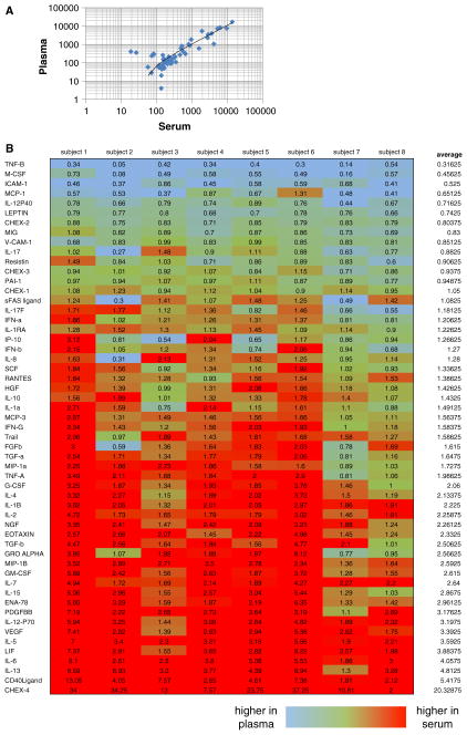
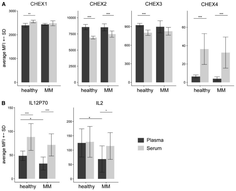
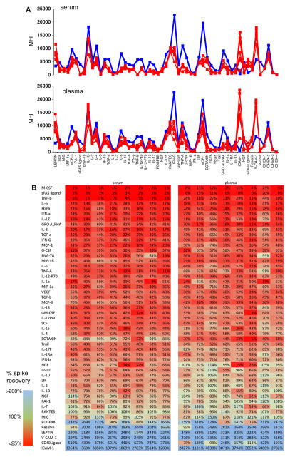
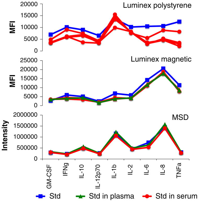
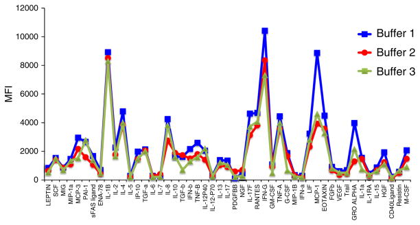
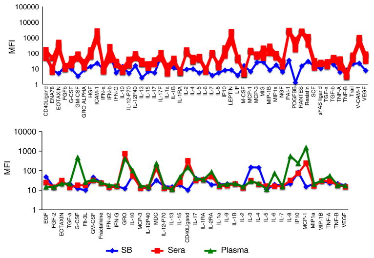
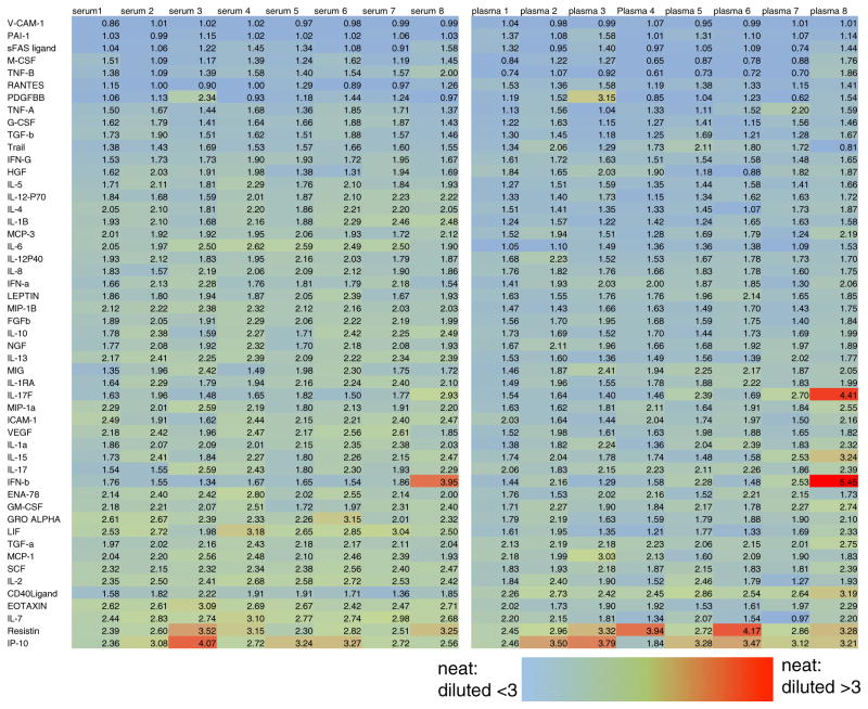
Multiplexed fluorescence or electrochemiluminescence immunoassays of soluble cytokines are commonly performed in the context of human serum or plasma, to look for disease biomarkers and to monitor the immune system in a simple and minimally invasive way. These assays provide challenges due to the complexities of the matrix (serum or plasma) and the presence of many cytokines near the limit of detection of the assay. Here, we compare the readout of matched serum and plasma samples, which are generally correlated. However, a subset of cytokines usually have higher levels in serum, and the non-specific background is significantly increased in serum versus plasma. Presumably as a result of this non-specific background, disease-related decreases in low-abundance cytokines can sometimes be detected in plasma but not in serum. We further show, through spike recovery experiments, that both serum and plasma inhibit the readout of many cytokines, with some variability between donors, but with serum causing greater inhibition than plasma in many cases. Standard diluents from different vendors can partially reverse this inhibition to varying degrees. Dilution of samples can also partly overcome the inhibitory effect of the matrix. We also show that dilution is nonlinear and differentially affects various cytokines. Together, these data argue that (1) plasma is a more sensitive matrix for detecting changes in certain low-abundance cytokines; (2) calculation of concentrations in serum or plasma matrices is inherently inaccurate; and (3) dilution of samples should not be assumed to be linear, i.e., all comparisons need to be made among similarly diluted samples.


Conflict of interest statement

Conflict of interest The authors declare that they have no conflict of interest.

Figures

Fig. 1
Fig. 1
a Correlation of median fluorescent intensity (MFI) of duplicate wells for plasma and serum samples from a representative healthy donor, for 51 cytokines analyzed in a single Luminex assay. Most cytokines are well correlated (ratio of plasma/serum values close to 1), with some spread at the low end of cytokine abundance and with a slightly higher average value for serum versus plasma. b Ratio of serum/plasma MFI for eight healthy donors analyzed for 51 cytokines in parallel in duplicate wells of serum or plasma. More cytokines show a higher value in serum than in plasma (red cells), but there is variability between donors
Fig. 2
Fig. 2
Plasma has a lower non-specific background and detects disease group differences that are not seen using serum. a CHEX control beads show similar average MFI between disease groups (healthy vs. MM). There are some significant differences between plasma and serum in CHEX1, 2, and 3. But most notably, CHEX 4, which assesses non-specific background, is significantly lower in plasma when compared to serum. b IL-12p70 and IL-2 show significantly higher plasma levels in healthy controls when compared to multiple myeloma patients. These differences are not significant in serum. IL-12p70 and IL-2 are low-level cytokines that decrease in disease groups relative to controls, and the lower non-specific background in plasma likely accounts for this. Significances were calculated using a two-sided Student’s t test. *p <0.05, **p <0.01, ***p <0.001. n = 6 (healthy), n = 10 (MM). MM = multiple myeloma
Fig. 3
Fig. 3
a Inhibitory effects of serum and plasma on cytokine standards. S6 standard (containing 625 pg/ml of each of 51 cytokines) was run under various conditions. Serum (top graph) or plasma (bottom graph) from eight healthy donors was used in place of the manufacturer’s standard dilution buffer (red lines), or the standard dilution buffer was used (blue lines). Inhibitory effects can be seen for many cytokines in the presence of serum or plasma, in that the red lines are significantly lower than the blue lines for both matrices. Inhibitory effects of serum appear to be somewhat more pronounced than plasma for certain cytokines, and there is some variability between different donors. b Heat map of the same data, showing the percent spike recovery of the S6 standard in the presence of serum or plasma from eight donors. Cytokines are ranked from worst (red) to best (blue) spike recovery. Many more cytokines have poor spike recovery than good; those with values >100 % reflect the quantitation of endogenous cytokine from the serum or plasma, in addition to the S6 standard (Color figure online)
Fig. 4
Fig. 4
Matrix effect in Luminex versus MesoScale Discovery (MSD) platforms. Cytokine standards were run using the manufacturer’s standard dilution buffer (blue squares) or with the buffer replaced by serum (red circles) or plasma (green triangles) of healthy donors. Standards contained 2,500 pg/ml (MSD and Luminex polystyrene bead kit) or 2,000 pg/ml (Luminex magnetic bead kit) of all cytokines (Color figure online)
Fig. 5
Fig. 5
Effect of different assay buffers on standard detection. Buffers intended for dilution of standards from MSD, Luminex (magnetic bead), and Luminex (polystyrene bead) kits were used to dilute the same S5 standard (representing 156.25 pg/ml of each of 51 cytokines). The buffers show generally similar patterns of cytokine detection, but the degree to which “Buffer 2” (MSD kit) and “Buffer 3” (Luminex magnetic bead kit) inhibit cytokine detection is greater than that for “Buffer 1” (Luminex polystyrene bead) in certain cases
Fig. 6
Fig. 6
Serum and plasma samples from healthy donors are sometimes lower than sample buffer (background). Serum (red squares) or plasma (green triangles) from several healthy donors was diluted 1:3 and run in a 51-plex polystyrene bead Luminex assay (Affymetrix, top panel) or a 39-plex magnetic bead Luminex assay (Millipore, bottom panel). MFI values are shown, compared with those of the sample buffer (blue diamonds). A few cytokines show significantly lower values in serum or plasma compared with the sample buffer (e.g., EGF, GM-CSF, IL-3, and IL-4) (Color figure online)
Fig. 7
Fig. 7
Dilution of serum or plasma is nonlinear. Heat map shows the ratio of MFI of neat/diluted (1:3 in PBS) plasma and serum samples from eight subjects. Most cytokines show a nonlinear dilution pattern, in which diluted samples have greater than 1/3 the MFI of neat samples. The effect may be slightly more pronounced in plasma and is also variable between donors (Color figure online)

References

    1. Djoba Siawaya JF, Roberts T, Babb C, Black G, Golakai HJ, Stanley K, Bapela NB, Hoal E, Parida S, van Helden P, Walzl G. An evaluation of commercial fluorescent bead-based Luminex cytokine assays. PLoS ONE. 2008;3:e2535. - PMC - PubMed
    1. duPont NC, Wang K, Wadhwa PD, Culhane JF, Nelson EL. Validation and comparison of luminex multiplex cytokine analysis kits with ELISA: determinations of a panel of nine cytokines in clinical sample culture supernatants. J Reprod Immunol. 2005;66:175–91. - PMC - PubMed
    1. Pang S, Smith J, Onley D, Reeve J, Walker M, Foy C. A comparability study of the emerging protein array platforms with established ELISA procedures. J Immunol Methods. 2005;302:1–12. - PubMed
    1. Breen EC, Reynolds SM, Cox C, Jacobson LP, Magpantay L, Mulder CB, Dibben O, Margolick JB, Bream JH, Sambrano E, Martinez-Maza O, Sinclair E, Borrow P, Landay AL, Rinaldo CR, Norris PJ. Multisite comparison of high-sensitivity multiplex cytokine assays. Clin Vaccine Immunol. 2011;18:1229–42. - PMC - PubMed
    1. Maecker HT. Measuring human cytokines. In: O’Gorman MR, Donnenberg AD, editors. Handbook of human immunology. 2. Boca Raton, FL: CRC Press; 2008. pp. 517–40.

Publication types