. 2014 Mar 15;192(6):2537-41.
doi: 10.4049/jimmunol.1303101. Epub 2014 Feb 12.

Cutting edge: IFN-γR signaling in non-T cell targets regulates T cell-mediated intestinal inflammation through multiple mechanisms

Affiliations

Cutting edge: IFN-γR signaling in non-T cell targets regulates T cell-mediated intestinal inflammation through multiple mechanisms

Jeong-su Do et al. J Immunol. .

Abstract

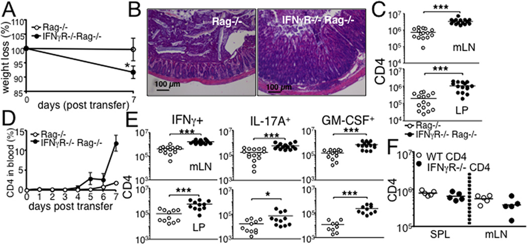
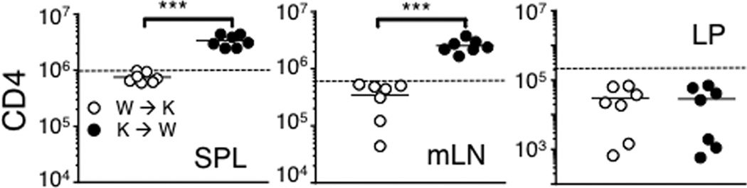
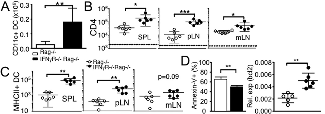
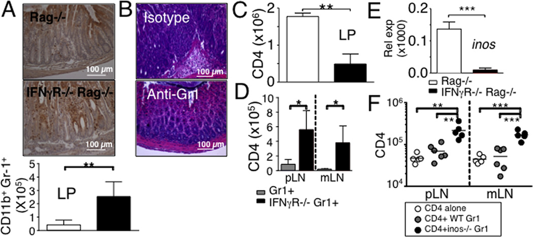
Naive CD4 T cells transferred into lymphopenic mice undergo spontaneous proliferation and induce chronic inflammation in the intestine. Cellular mechanisms regulating the proliferative and inflammatory processes are not fully understood. In this study, we report that IFN-γ signaling in host cells plays a major role in limiting both T cell expansion and T cell-induced intestinal inflammation. However, the role of IFN-γ appears to differ depending on the target cells. IFN-γ signaling in dendritic cells controls T cell expansion, whereas IFN-γ signaling in neutrophils seems to regulate both T cell expansion and inflammation. IFN-γ signaling in nonhematopoietic cells may control inflammation. Therefore, our results suggest novel immunoregulatory functions for IFN-γ to orchestrate colitogenic T cell responses through its distinct action on different non-T cell target cells.


Figures

Figure 1
Figure 1. Rag−/− mice deficient in IFNγR develop severe colon inflammation following naïve CD4 T cell transfer
FACS purified naive Thy1.1+ CD4 T cells were transferred into Rag−/− or IFNγR−/− Rag−/− recipients. (A) Weight loss after T cell transfer. (B) The colon tissues were harvested 7 days post transfer and H&E stained. 20x magnification. (C) Absolute numbers of total donor CD4 T cells in the indicated tissues were enumerated 7 days post transfer by FACS analysis. (D) The recipients were daily bled, and circulating donor T cells were examined. (E) Cells from the indicated tissues were harvested 7 days post transfer, ex vivo stimulated with PMA/Ionomycin, and stained for intracellular cytokine expression. Absolute numbers of IFNγ, IL-17A and GM-CSF producing CD4 T cells were determined by FACS analysis. (F) CD4 T cells isolated from Thy1.1 WT or IFNγR−/− mice were transferred into Rag−/− recipients. The absolute numbers of the donor T cells was examined 7 days post transfer. All experiments were repeated 2-3 times and similar results were observed. Each symbol represents individually tested animals. *, p<0.05; ***, p<0.001.
Figure 2
Figure 2. IFNγ signaling in hematopoietic cells is essential to control CD4 T cell expansion
Lethally irradiated IFNγR−/− Rag−/− and Rag−/− mice were reconstituted with BM cells from Rag−/− and IFNγR−/− Rag−/− donor mice, respectively (Rag−/− BM → IFNγR−/− Rag−/−, W → K and IFNγR−/− Rag−/− BM → Rag−/−, K → W). FACS purified naive Thy1.1+ CD4 T cells were transferred into the reconstituted recipients after 6 weeks of BM reconstitution. The T cell recipients were sacrificed 7 days post transfer, and cells from the indicated tissues were stained for Thy1.1 and CD4. Absolute numbers of donor cells were calculated. T cell recovery of unmanipulated Rag−/− recipients is shown as dotted line. All experiments were repeated twice and similar results were observed. Each symbol represents individually tested animals. ***, p<0.001.
Figure 3
Figure 3. Prolonged IFNγR−/− DC survival promotes CD4 T cell expansion
(A) The absolute numbers of CD11c+ DCs in the colon was calculated 7 days post transfer. Data are the mean ± SD of 5-6 individually tested mice from two separate experiments. (B) MHCII−/− Rag−/− mice received FACS purified Ly5.1+ naive CD4 T cells together with splenic CD11c+ DCs isolated from WT (○) or IFNγR−/− (●) mice. Donor CD4 T cell recovery was determined by FACS 7 days post transfer. (C) Absolute numbers of adoptively transferred WT and IFNγR−/− MHCII+ CD11c+ DCs were counted at day 7. All the experiments were repeated twice and similar results were observed. (D) Annexin-V staining and bcl2 expression of purified mLN DCs were examined. Data are the mean ± SD of 5-6 individually tested mice. *, p<0.05; **, p<0.01; ***, p<0.001.
Figure 4
Figure 4. Gr1+ cells in response to IFNγ regulate CD4 T cells expansion and intestinal inflammation in IFNγR−/− Rag−/− mice by iNOS-dependent mechanism
(A) Immunohistochemistry analysis of the Gr1+ cells in the colon. Absolute numbers of Gr1+ CD11b+ cells in the colon were determined 7 days post naïve CD4 T cell transfer. Data shown represent the mean ± SD of 6 individually tested mice from two independent experiments. (B and C) IFNγR−/− Rag−/− recipients of naïve CD4 T cells were treated with RB6-8C5 anti-Gr1 mAb (150 µg) at days -1, 2, and 5 of T cell transfer. H&E staining of the colon tissues and the absolute numbers of CD4 T cells in the colon were determined at 7 days post transfer. (D) Thy1.1 naive CD4 T cells were transferred into Rag−/− recipients together with Gr1+ cells harvested from WT or IFNγR−/− mice. Donor T cells recovery was measured at day 7. (E) iNOS expression in the colon was examined by real time PCR at 7 days post CD4 transfer. (F) Naive CD4 T cells were transferred into Rag−/− recipients together with Gr1+ cells harvested from WT or iNOS−/− mice. Donor T cells recovery was measured at day 7. Data are the mean ± SD of 4-9 individually tested animals from 2-3 independent experiments. *, p<0.05; **, p<0.01; ***, p<0.001.

References

    1. Powrie F, Leach MW, Mauze S, Menon S, Caddle LB, Coffman RL. Inhibition of Th1 responses prevents inflammatory bowel disease in scid mice reconstituted with CD45RBhi CD4+ T cells. Immunity. 1994;1:553–562. - PubMed
    1. Ostanin DV, Bao J, Koboziev I, Gray L, Robinson-Jackson SA, Kosloski-Davidson M, Price VH, Grisham MB. T cell transfer model of chronic colitis: concepts, considerations, and tricks of the trade. American journal of physiology. Gastrointestinal and liver physiology. 2009;296:G135–G146. - PMC - PubMed
    1. Do JS, Visperas A, Dong C, Baldwin WM, 3rd, Min B. Cutting edge: Generation of colitogenic Th17 CD4 T cells is enhanced by IL-17+ gammadelta T cells. J Immunol. 2011;186:4546–4550. - PMC - PubMed
    1. Do JS, Foucras G, Kamada N, Schenk AF, Shaw M, Nunez G, Paul WE, Min B. Both exogenous commensal and endogenous self antigens stimulate T cell proliferation under lymphopenic conditions. Cellular immunology. 2012;272:117–123. - PMC - PubMed
    1. Do JS, Min B. Differential requirements of MHC and of DCs for endogenous proliferation of different T-cell subsets in vivo. Proc Natl Acad Sci U S A. 2009;106:20394–20398. - PMC - PubMed

Publication types

MeSH terms

Substances