. 2014 Feb 12;34(7):2438-43.
doi: 10.1523/JNEUROSCI.2947-13.2014.

Astroglial-derived periostin promotes axonal regeneration after spinal cord injury

Affiliations

Astroglial-derived periostin promotes axonal regeneration after spinal cord injury

Chung-Hsuan Shih et al. J Neurosci. .

Abstract

Traumatic spinal cord injury (SCI) results in a cascade of tissue responses leading to cell death, axonal degeneration, and glial scar formation, exacerbating the already hostile environment and further inhibiting axon regeneration. Overcoming these inhibitory cues and promoting axonal regeneration is one of the primary targets in developing a cure for SCI. Previously, we demonstrated that transplantation of bone morphogenetic protein (BMP)-induced astrocytes derived from embryonic glial-restricted precursors (GDAs(BMP)) promotes extensive axonal growth and motor function recovery in a rodent spinal cord injury model. Here, we identify periostin (POSTN), a secreted protein, as a key component of GDA(BMP)-induced axonal regeneration. POSTN is highly expressed by GDAs(BMP) and the perturbation of POSTN expression by shRNA diminished GDA(BMP)-induced neurite extension in vitro. We also found that recombinant POSTN is sufficient to overcome the inhibitory effect of scar-associated molecules and promote neurite extension in vitro by signaling through focal adhesion kinase and Akt. Furthermore, transplantation of POSTN-deficient GDAs(BMP) into the injured rat spinal cord resulted in compromised axonal regeneration, indicating that POSTN plays an essential role in GDA(BMP)-mediated axonal regeneration. This finding reveals not only one of the major mechanisms underlying GDA(BMP)-dependent recovery from SCI, but also the potential of POSTN as a therapeutic agent for traumatic injury of the CNS.

Keywords: astrocyte; cell therapy; glial precursor; neurite outgrowth; spinal cord; support cell.


Figures

Figure 1.
Figure 1.
POSTN mediates GDABMP-induced neurite extension in vitro. a, Adult DRGNs were cultured in conditioned medium (CM) from GRPs, GDAsBMP, or GDAsCNTF. After 3 d, DRGNs were immunostained with TujI antibody. Scale bar, 100 μm. Relative axon length of DRGNs grown in neurobasal medium (NB), CM from GRPs, GDAsBMP, or GDAsCNTF. (n = 3). Data are mean ± SD. *p < 0.05, t test. Expression level of POSTN in GRP, GDAsBMP, and GDAsCNTF measured by semiquantitative PCR (b) or Western blot analysis (c). d, GRP cells stably transduced with vector or three different POSTN-specific shRNA expression constructs. GRPs were either maintained as precursors or differentiated into GDAsBMP. POSTN was detected in CM and lysates by Western blot. e, DRGNs cultured for 3 d in CM collected from GRPs or GDAsBMP infected with either control or POSTN shRNA constructs. Upper panels show representative images of cultured DRGs. Lower panel presents quantification of relative neurite lengths. Neurites labelled by TujI immunostaining. Scale bar, 100 μm. Quantitative result of e (n = 3). Data are mean ± SD. *p < 0.05, t test.
Figure 2.
Figure 2.
Recombinant POSTN overcomes inhibitory effect of myelin and chondroitin sulfate proteoglycans and promotes neurite outgrowth in vitro through FAK and Akt signaling pathway. a, b, Adult DRGNs (a) and P7 CGNs (b) cultured with or without myelin/CSPGs in the presence or absence of recombinant POSTN. Cells were immunostained with TujI antibody. Scale bars, 100 μm. Mean neurite length is shown in e (n = 3). Data are mean ± SD. *p < 0.05, t test. c, CGNs treated with control vehicle or recombinant POSTN 4 h after plating. Cell lysates were harvested 2 and 16 h after treatment for Western blot analysis. Relative expression levels were normalized to b-actin and then untreated samples at 2 h. d, CGNs were seeded with or without recombinant POSTN and treated with control vehicle or FAK/Akt inhibitors. Relative axon length 24 h after plating is shown (n = 3). Data are mean ± SD. *p < 0.05, ANOVA.
Figure 3.
Figure 3.
POSTN is highly expressed in GDABMP, but not in postnatal astrocytes or adult spinal cord. a, GRPs or postnatal O2As were either maintained as precursors or differentiated into GDAsBMP, GDAsCNTF, type 2 astrocytes (2-A), or preoligodendrocytes by exposing to BMP or CNTF, respectively. Expression level of POSTN was measured by semiquantitative PCR. b, Embryonic GRPs and postnatal astrocytes cultured in vitro for 20, 40, or 60 d were treated with FGF, BMP, or CNTF for 6 d. Expression of POSTN was measured by Western blot analysis. c, Expression level of POSTN in GRPs, GDAsBMP, intact (distal) or injured (center) adult spinal cord tissues measured by Western blot analysis. Expression of the inflammatory protein HO-1 maps the injury.
Figure 4.
Figure 4.
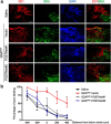
POSTN mediates GDABMP-dependent axonal regeneration after SCI. Animals receiving hemisection SCI injury placed on right side of dorsal column at C1/2 treated with saline or GDAsBMP infected with control vector or POSTN-shRNA vector. Biotinylated dextran amine was injected at the C3/4 level of the dorsal column on the injured side after injury/transplantation and animals were killed 7 d later. a, Sagittal sections of immunostained spinal cord (ED1 and anterograde axonal tracing by BDA). Nuclei were counterstained with DAPI. Scale bar, 200 μm. b, Percentage of labeled axons growing into lesion center (n = 4). Data are mean ± SEM. *p < 0.05, ANOVA.

References

    1. Bao S, Ouyang G, Bai X, Huang Z, Ma C, Liu M, Shao R, Anderson RM, Rich JN, Wang XF. Periostin potently promotes metastatic growth of colon cancer by augmenting cell survival via the Akt/PKB pathway. Cancer Cell. 2004;5:329–339. doi: 10.1016/S1535-6108(04)00081-9. - DOI - PubMed
    1. Chen J, Joon Lee H, Jakovcevski I, Shah R, Bhagat N, Loers G, Liu HY, Meiners S, Taschenberger G, Kügler S, Irintchev A, Schachner M. The extracellular matrix glycoprotein tenascin-C is beneficial for spinal cord regeneration. Mol Ther. 2010;18:1769–1777. doi: 10.1038/mt.2010.133. - DOI - PMC - PubMed
    1. Davies JE, Huang C, Pröschel C, Noble M, Mayer-Pröschel M, Davies SJ. Astrocytes derived from glial-restricted precursors promote spinal cord repair. J Biol. 2006;5:7. doi: 10.1186/jbiol35. - DOI - PMC - PubMed
    1. Davies JE, Pröschel C, Zhang N, Noble M, Mayer-Pröschel M, Davies SJ. Transplanted astrocytes derived from BMP- or CNTF-treated glial-restricted precursors have opposite effects on recovery and allodynia after spinal cord injury. J Biol. 2008;7:24. doi: 10.1186/jbiol85. - DOI - PMC - PubMed
    1. Davies SJ, Shih CH, Noble M, Mayer-Pröschel M, Davies JE, Pröschel C. Transplantation of specific human astrocytes promotes functional recovery after spinal cord injury. PLoS One. 2011;6:e17328. doi: 10.1371/journal.pone.0017328. - DOI - PMC - PubMed

Publication types