. 2014 Feb 12;34(7):2503-13.
doi: 10.1523/JNEUROSCI.2830-13.2014.

Trans-signaling is a dominant mechanism for the pathogenic actions of interleukin-6 in the brain

Affiliations

Trans-signaling is a dominant mechanism for the pathogenic actions of interleukin-6 in the brain

Iain L Campbell et al. J Neurosci. .

Abstract

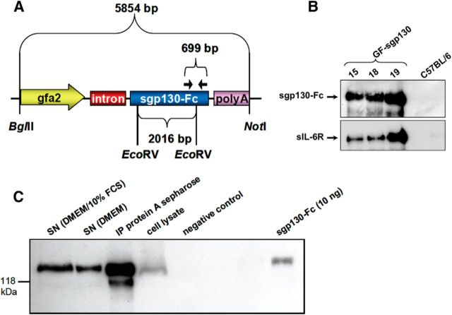
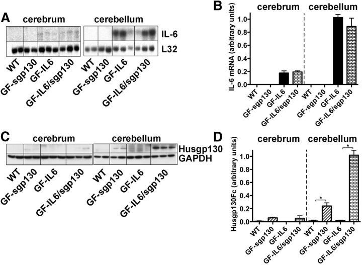
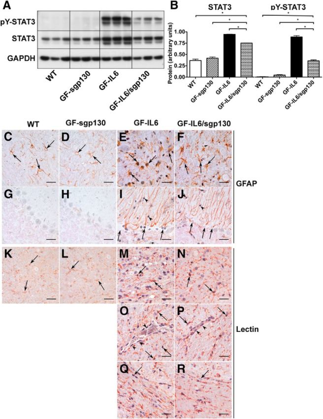
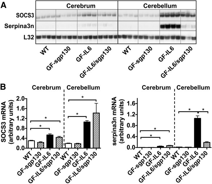
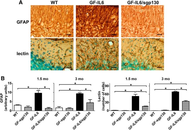
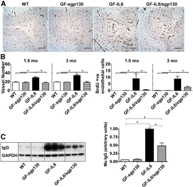
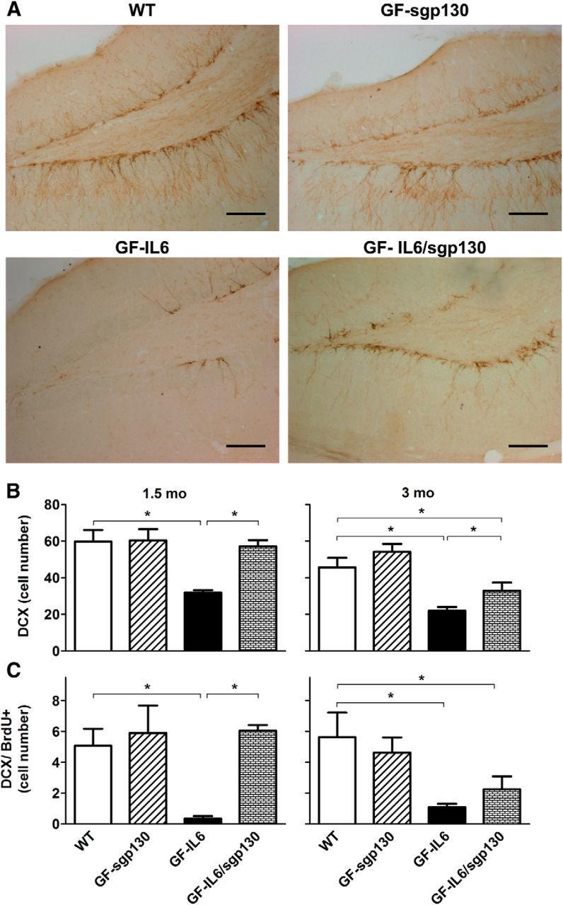
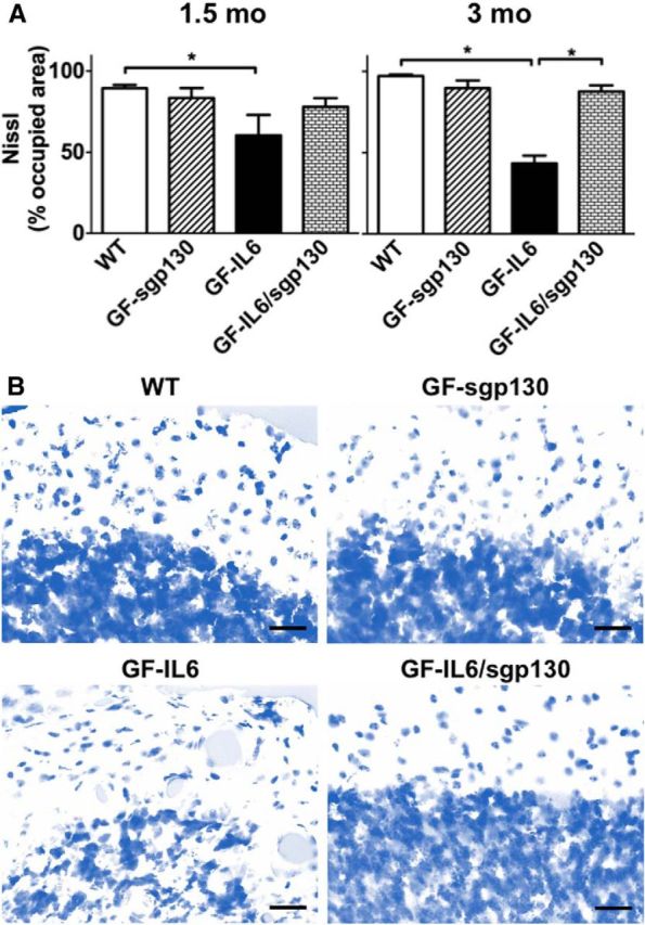
IL-6 is implicated in the pathogenesis of various neuroinflammatory and neurodegenerative disorders of the CNS. IL-6 signals via binding to either the membrane bound IL-6Rα (classic signaling) or soluble (s)IL-6Ra (trans-signaling) that then form a complex with gp130 to activate the JAK/STAT signaling pathway. The importance of classic versus trans-signaling in mediating IL-6 actions in the living CNS is relatively unknown and was the focus of this investigation. Bigenic mice (termed GFAP-IL6/sgp130 mice) were generated with CNS-restricted, astrocyte-targeted production of IL-6 and coproduction of the specific inhibitor of IL-6 trans-signaling, human sgp130-Fc. Transgene-encoded IL-6 mRNA levels were similar in the brain of GFAP-IL6 and GFAP-IL6/sgp130 mice. However, GFAP-IL6/sgp130 mice had decreased pY(705)-STAT3 in the brain due to a reduction in the total number of pY(705)-STAT3-positive cells and a marked loss of pY(705)-STAT3 in specific cell types. Blockade of trans-signaling in the brain of the GFAP-IL6 mice significantly attenuated Serpina3n but not SOCS3 gene expression, whereas vascular changes including angiogenesis and blood-brain barrier leakage as well as gliosis were also reduced significantly. Hippocampal neurogenesis which was impaired in GFAP-IL6 mice was rescued in young GFAP-IL6 mice with cerebral sgp130 production. Finally, degenerative changes in the cerebellum characteristic of GFAP-IL6 mice were absent in GFAP-IL6/sgp130 mice. The findings indicate that in the CNS: (1) sgp130 is able to block IL-6 trans-signaling, (2) trans-signaling is important for IL-6 cellular communication with selective cellular and molecular targets, and (3) blocking of trans-signaling alleviates many of the detrimental effects of IL-6.

Keywords: interleukin-6; neuropathology; signal transduction; trans-signaling; transgenic mouse.


Figures

Figure 1.
Figure 1.
Generation and characterization of GFAP-sgp130 transgenic mice. A, Schematic representation of the organization of the gfa2-sgp130-Fc optimized cDNA expression cassette. B, Immunoblot detection of secreted sgp130-Fc from primary astrocytes of GFAP-sgp130 transgenic mice or WT controls. Cell culture supernatants (SNs) were used directly or after coprecipitation of sgp130-Fc with protein A Sepharose. C, Immunoblot analysis after coprecipitation of hyper-IL-6 with brain-derived sgp130-Fc from GFAP-sgp130 transgenic mice. Brain homogenates from the three independent transgenic mouse lines 15, 18, and 19 or from WT mice were incubated with hyper-IL-6 and coprecipitated with protein A Sepharose. Coprecipitated hyper-IL-6 was detected with an IL-6R monoclonal (clone 14-18), whereas sgp130-Fc was detected with the gp130 antibody B-P4. Recombinant sgp130-Fc (10 ng) was included as positive control.
Figure 2.
Figure 2.
Transgene-encoded IL-6 mRNA and human sgp130 in the brain of GFAP-IL6 and GFAP-IL6/sgp130 mice. A, Total RNA was extracted from the brain of 1.5-month-old mice and analyzed (5 μg per lane) by RPA and visualized by autoradiography. B, Quantification of the band densities in A for IL-6 relative to the housekeeping control L32 was performed by densitometry using NIH ImageJ software. Values represent the mean ± SEM for two experiments with n = 6 independent samples; *p ≤ 0.05. C, Tissue lysates (20 μg protein per lane) from brain of 1.5-month-old mice were subjected to SDS-PAGE followed by immunoblotting. D, Quantification of the band densities in C for sgp130 relative to GAPDH was performed by densitometry using NIH ImageJ software. Values represent the mean ± SEM for two experiments with n = 4 or 6 independent samples; *p ≤ 0.05.
Figure 3.
Figure 3.
The presence of sgp130 reduces steady-state pY-STAT3 in the cerebellum and in specific cells of the cerebellum of GFAP-IL6 mice. A, Tissue lysates (20 μg protein per lane) from cerebellum of 1.5-month-old mice were subjected to SDS-PAGE followed by immunoblotting. B, Quantification of the band densities in A for pY-STAT3 or STAT3 relative to GAPDH was performed by densitometry using NIH ImageJ software. Values represent the mean ± SEM with n = 3 brains per genotype; *p ≤ 0.05. CR, Paraformaldehyde fixed, paraffin-embedded brain sections (5 μm) prepared from 3-month-old mice were processed for immunohistochemistry for pY-STAT3 (purple) and combined with immunohistochemistry for GFAP (red) or histochemistry using tomato lectin binding (red). Scale bar, 25 μm.
Figure 4.
Figure 4.
Inhibition of IL-6 trans-signaling resulted in differential expression of the Socs3 versus Serpina3n genes in the cerebellum of GFAP-IL6 mice. A, Total RNA was extracted from the brain of 1.5-month-old mice and analyzed (5 μg per lane) by RPA and visualized by autoradiography. B, Quantification of the band densities in A for SOCS3 or Serpina3n relative to the housekeeping control L32 was performed by densitometry using NIH ImageJ software. Values represent the mean ± SEM with n = 3 or 4 brains per genotype; *p ≤ 0.05.
Figure 5.
Figure 5.
IL-6 trans-signaling contributed to the development of gliosis in the cerebellum of GFAP-IL6 mice. A, Paraformaldehyde fixed, free-floating brain sections (30 μm) prepared from 1.5-month-old mice were processed for immunohistochemical detection of GFAP or lectin histochemistry. Scale bar, 25 μm. B, Staining in A and for similarly stained brain sections from 3-month-old mice were quantified with the NIH ImageJ analysis software. These analyses were performed on at least three blinded sections per brain and on a minimum of three brains per genotype. Values represent the mean ± SEM; *p ≤ 0.05.
Figure 6.
Figure 6.
Abnormalities in vascular structure, proliferation and function were reduced significantly in the cerebellum of GFAP-IL6 mice by the inhibition of IL-6 trans-signaling. A, Laminin immunohistochemistry was performed on paraformaldehyde fixed, paraffin-embedded sections (5 μm) of brain prepared from 3-month-old mice. Scale bar, 50 μm. B, Vasculogenesis was assessed in free-floating brain sections (30 μm) and quantified by direct counting of lectin-stained blood vessels (left) or BrdU-positive endothelial cells (right). This analysis was performed on at least three blinded sections per brain and on a minimum of three brains per genotype. Values represent the mean ± SEM; *p ≤ 0.05. C, The degree of BBB leakage was determined by quantifying the levels of IgG present in the cerebellum of 3-month-old mice. Tissue lysates (20 μg protein per lane) were prepared and subject to SDS-PAGE followed by immunoblotting for murine IgG. Quantification of the band densities in C for IgG relative to GAPDH was performed by densitometry using NIH ImageJ software. Values represent the mean ± SEM with n = 3 or 4 brains per genotype; *p ≤ 0.05.
Figure 7.
Figure 7.
Defective hippocampal neurogenesis was rescued in young GFAP-IL6 mice with IL-6 trans-signaling blockade. A, Paraformaldehyde fixed, free-floating brain sections (30 μm) prepared from 1.5- or 3-month-old mice that were injected with BrdU, were processed for immunohistochemical detection of DCX. Scale bar, 100 μm. B, Quantification of the data presented in A. This analysis was performed on at least three blinded sections per brain and on a minimum of three brains per genotype. Values represent the mean ± SEM; *p ≤ 0.05. C, A separate set of free-floating brain sections was processed for dual-label immunohistochemical detection of DCX and BrdU and the number of DCX and BrdU double-positive cells counted. This analysis was performed on at least three blinded sections per brain and on a minimum of three brains per genotype. Values represent the mean ± SEM; *p ≤ 0.05.
Figure 8.
Figure 8.
Inhibition of IL-6 trans-signaling prevents neurodegeneration in the cerebellum of GFAP-IL6 mice. A, Free-floating brain sections were stained with Nissl and quantified with the NIH ImageJ analysis software. This analysis was performed on at least three blinded sections per brain and on a minimum of three brains per genotype. Values represent the mean ± SD; *p ≤ 0.05. B, Nissl stained free-floating brain sections (30 μm) from 3-month-old mice. Note loss of architecture of the granule and molecular cell layers, as well as loss of cellularity in GFAP-IL6 cerebellum compared with an age-matched GFAP-IL6/sgp130 littermate. Scale bar, 25 μm.

References

    1. Auernhammer CJ, Bousquet C, Melmed S. Autoregulation of pituitary corticotroph SOCS-3 expression: characterization of the murine SOCS-3 promoter. Proc Natl Acad Sci U S A. 1999;96:6964–6969. doi: 10.1073/pnas.96.12.6964. - DOI - PMC - PubMed
    1. Brett FM, Mizisin AP, Powell HC, Campbell IL. Evolution of neuropathologic abnormalities associated with blood–brain barrier breakdown in transgenic mice expressing interleukin-6 in astrocytes. J Neuropathol Exp Neurol. 1995;54:766–775. doi: 10.1097/00005072-199511000-00003. - DOI - PubMed
    1. Brunello AG, Weissenberger J, Kappeler A, Vallan C, Peters M, Rose-John S, Weis J. Astrocytic alterations in interleukin-6/soluble interleukin-6 receptor alpha double-transgenic mice. Am J Pathol. 2000;157:1485–1493. doi: 10.1016/S0002-9440(10)64787-6. - DOI - PMC - PubMed
    1. Burton MD, Sparkman NL, Johnson RW. Inhibition of interleukin-6 trans-signaling in the brain facilitates recovery from lipopolysaccharide-induced sickness behavior. J Neuroinflammation. 2011;8:54. doi: 10.1186/1742-2094-8-54. - DOI - PMC - PubMed
    1. Burton MD, Rytych JL, Freund GG, Johnson RW. Central inhibition of interleukin-6 trans-signaling during peripheral infection reduced neuroinflammation and sickness in aged mice. Brain Behav Immun. 2013;30:66–72. doi: 10.1016/j.bbi.2013.01.002. - DOI - PMC - PubMed

Publication types