Biodegradable and thermosensitive micelles inhibit ischemia-induced postoperative peritoneal adhesion
- PMID: 24523585
- PMCID: PMC3921091
- DOI: 10.2147/IJN.S55497
Biodegradable and thermosensitive micelles inhibit ischemia-induced postoperative peritoneal adhesion
Abstract
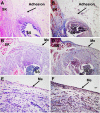
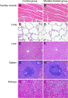
Ischemia-induced adhesion is very common after surgery, and leads to severe abdominal adhesions. Unfortunately, many existing barrier agents used for adhesion prevention have only limited success. The objective of this study is to evaluate the efficacy of biodegradable and thermosensitive poly(ε-caprolactone)-poly(ethylene glycol)-poly(ε-caprolactone) (PCL-PEG-PCL) micelles for the prevention of postoperative ischemia-induced adhesion. We found that the synthesized PCL-PEG-PCL copolymer could self-assemble in an aqueous solution to form micelles with a mean size of 40.1 ± 2.7 nm at 10°C, and the self-assembled micelles could instantly turn into a nonflowing gel at body temperature. In vitro cytotoxicity tests suggested that the copolymer showed little toxicity on NIH-3T3 cells even at amounts up to 1,000 μg/mL. In the in vivo test, the postsurgical ischemic-induced peritoneal adhesion model was established and then treated with the biodegradable and thermosensitive micelles. In the control group (n=12), all animals developed adhesions (mean score, 3.58 ± 0.51), whereas three rats in the micelles-treated group (n=12) did not develop any adhesions (mean score, 0.67 ± 0.78; P<0.001, Mann-Whitney U-test). Both hematoxylin and eosin and Masson trichrome staining of the ischemic tissues indicated that the micelles demonstrated excellent therapeutic effects on ischemia-induced adhesion. On Day 7 after micelle treatment, a layer of neo-mesothelial cells emerged on the injured tissues, which confirmed the antiadhesion effect of the micelles. The thermosensitive micelles had no significant side effects in the in vivo experiments. These results suggested that biodegradable and thermosensitive PCL-PEG-PCL micelles could serve as a potential barrier agent to reduce the severity of and even prevent the formation of ischemia-induced adhesions.
Keywords: poly(ε-caprolactone)–poly(ethylene glycol)–poly(ε-caprolactone) (PCL–PEG–PCL); postsurgical adhesion; surgical complications, barrier agent.
Figures


Comment in
-
Challenging nanoparticles: a target of personalized adhesion prevention strategy.Int J Nanomedicine. 2014 Jul 15;9:3375-6. doi: 10.2147/IJN.S67938. eCollection 2014. Int J Nanomedicine. 2014. PMID: 25075184 Free PMC article. No abstract available.
References
-
- Gong CY, Wu QJ, Liao JF, et al. Prevention of postsurgical cauterization-induced peritoneal adhesions by biodegradable and thermosensitive micelles. J Biomed Nanotechnol. 2013;9(12):1984–1995. - PubMed
-
- Ahn HS, Lee H-J, Yoo M-W, et al. Efficacy of an injectable thermosensitive gel on postoperative adhesion in rat model. J Korean Surg Soc. 2010;79(4):239–245.
-
- Liakakos T, Thomakos N, Fine PM, Dervenis C, Young RL. Peritoneal adhesions: etiology, pathophysiology, and clinical significance. Recent advances in prevention and management. Dig Surg. 2001;18(4):260–273. - PubMed
Publication types
MeSH terms
Substances
LinkOut - more resources
Full Text Sources
Other Literature Sources
Medical

