Review
. 2014 Jan 30:8:5.
doi: 10.3389/fnsys.2014.00005. eCollection 2014.

The thalamostriatal system in normal and diseased states

Affiliations
Review

The thalamostriatal system in normal and diseased states

Yoland Smith et al. Front Syst Neurosci. .

Abstract

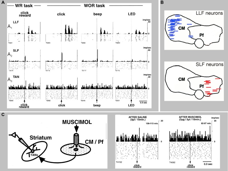
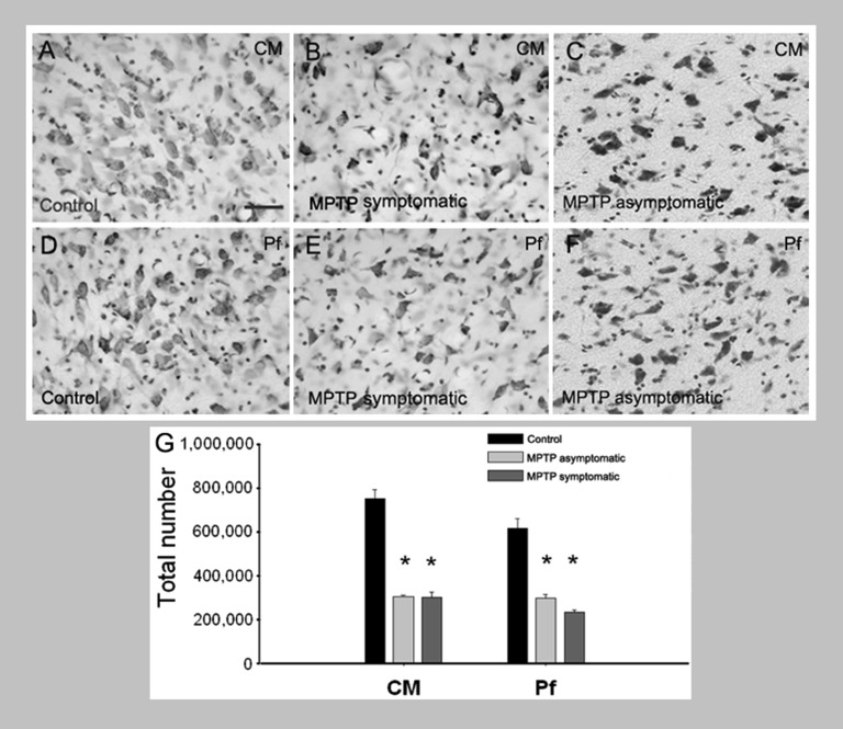
Because of our limited knowledge of the functional role of the thalamostriatal system, this massive network is often ignored in models of the pathophysiology of brain disorders of basal ganglia origin, such as Parkinson's disease (PD). However, over the past decade, significant advances have led to a deeper understanding of the anatomical, electrophysiological, behavioral and pathological aspects of the thalamostriatal system. The cloning of the vesicular glutamate transporters 1 and 2 (vGluT1 and vGluT2) has provided powerful tools to differentiate thalamostriatal from corticostriatal glutamatergic terminals, allowing us to carry out comparative studies of the synaptology and plasticity of these two systems in normal and pathological conditions. Findings from these studies have led to the recognition of two thalamostriatal systems, based on their differential origin from the caudal intralaminar nuclear group, the center median/parafascicular (CM/Pf) complex, or other thalamic nuclei. The recent use of optogenetic methods supports this model of the organization of the thalamostriatal systems, showing differences in functionality and glutamate receptor localization at thalamostriatal synapses from Pf and other thalamic nuclei. At the functional level, evidence largely gathered from thalamic recordings in awake monkeys strongly suggests that the thalamostriatal system from the CM/Pf is involved in regulating alertness and switching behaviors. Importantly, there is evidence that the caudal intralaminar nuclei and their axonal projections to the striatum partly degenerate in PD and that CM/Pf deep brain stimulation (DBS) may be therapeutically useful in several movement disorders.

Keywords: Parkinson’s disease; Tourette’s syndrome; attention; glutamate; intralaminar nuclei; striatum; thalamus; vesicular glutamate transporter.


Figures

Figure 1
Figure 1
Summary of the anatomical, functional and pathological characteristics that differentiate thalamostriatal projections from the CM/Pf vs. other thalamic nuclei (i.e., non CM/Pf).
Figure 2
Figure 2
Differential pattern of activity and synaptic connectivity of CM/Pf vs. non-CM/Pf thalamostriatal terminals. (A) Thalamostriatal neurons of the central lateral nucleus (non-CM/Pf) have a compact bushy dendritic arbor. They tend to fire brief bursts of spikes at high frequency (2–5 spikes at ~150 Hz) in time with the active part of the cortical slow oscillation, reminiscent of low-threshold Ca2+ spike bursts. At the ultrastructural level, terminals from the central lateral nucleus almost exclusively target spines in the striatum. (B) Thalamostriatal neurons of the parafascicular nucleus have a reticular dendritic arbor with long and infrequently branching dendrites. They also tend to fire bursts of action potentials in time with the active part of the cortical slow oscillation, but the burst spike frequency is significantly lower and more variable than those of central lateral neurons. At the ultrastructural level, terminals from the parafascicular nucleus predominantly target the dendritic shafts of striatal neurons (see Lacey et al., , for more details). (C) Histogram showing the frequency of axo-dendritic vs. axo-spinous synapses formed by anterogradely labeled boutons from various thalamic nuclei and the primary motor cortex (M1) in the rat striatum. Note the striking difference in the pattern of synaptic connectivity of terminals from Pf vs. other thalamic nuclei (see Raju et al., for more details). Abbreviations: AV: Anteroventral nucleus; CL: Central lateral nucleus; LD: Laterodorsal nucleus; MD: Mediodorsal nucleus; M1: Primary motor cortex; Pf: Parafascicular nucleus; VA/VL: Ventral anterior/ventral lateral nucleus.
Figure 3
Figure 3
Electrophysiological responses of striatal neurons to electrical stimulation of CM in awake monkeys. Electrical stimulation (100 Hz, 100 pulses-shaded area) of CM evokes responses in PANs (putatively MSNs) and TANs (putatively cholinergic interneurons) in awake rhesus monkeys. (A) Example of a PAN responding with increased firing to electrical CM stimulation. (B) Example of a TAN responding with a brief decrease followed by an increase in firing to CM stimulation. The histograms and rasters are aligned to the start of stimulation trains. (C) Summary of responses. The majority of PANs display increases or decreases in firing rate, while most TANs present combinatory (increases and decreases) responses following CM stimulation (see Nanda et al., for more details).
Figure 4
Figure 4
Electrophysiological responses of striatal MSNs to optogenetic activation of thalamostriatal terminals from CL or Pf in mice. (A) Channelrhodopsin-2 (ChR2) was delivered to either the CL or Pf thalamic nucleus using a stereotaxic injection of adeno-associated virus containing the double-floxed sequence for ChR2-YFP in CAMKII-cre mice. This approach enabled expression of ChR2 in the excitatory thalamic neurons of either the CL or Pf nucleus including in their axonal arbor. Thalamic axons expressing ChR2-YFP were readily visible in acute striatal slices and this method allowed for activation of only those synapses originating from neurons from the injected thalamic nucleus by illumination of these slices with blue (473 nm) laser or LED light. (B) Whole-cell patch-clamp recordings of striatal MSNs showed that activation of CL synapses led to consistently larger post-synaptic responses than activation of Pf synapses. (C) Detailed investigation of the glutamate receptor-mediated currents revealed the CL synapses exhibit predominantly AMPA receptor-mediated currents in response to light activation and the Pf synapses exhibit predominantly NMDA receptor-mediated currents. (D) The short term plastic properties of these synapses were investigated by repetitive activation of the synapses in close succession. This revealed that inputs from CL exhibit short term facilitation, in which the response to the second activation has a larger amplitude than the response to the first activation, whereas inputs from Pf exhibit short term depression. (E) The long term plastic properties of these synapses were investigated using a spike timing-dependent plasticity protocol, consisting of the pairing of optical activation of a pre-synaptic thalamic input with a post-synaptic action potential in a MSN. This protocol induced a clear long term depression of synaptic efficacy at Pf synapses, but no plasticity was observed at CL synapses. The same observation was made using either a pre-post or post-pre pairing, with only the pre-post pairing shown for clarity (see Ellender et al., for more details).
Figure 5
Figure 5
Sensory responses of two types of CM/Pf neurons, and a striatal TAN recorded during the presentation of a stimulus with or without reward (WR vs. WOR). The spike rasters and histograms are aligned to the time of presentation of the stimulus. (A1) Representative activity of a CM neuron with long-latency facilitation (LLF) following stimulus presentation. (A2) Activity of a Pf neuron showing short-latency facilitation (SLF) after stimulus. (A3) Activity of a TAN. Note that thalamic responses occur in both WR and WOR tasks, whereas TAN responses occur only in the WR task. (B) Localization of LLF and SLF neurons in the monkey CM/Pf complex. Note the complete segregation of these two populations of neurons in CM or Pf. (C) Application of the GABAA receptor agonist muscimol in CM/Pf alters the pattern of responses of striatal TANs to the presentation of reward-related sensory stimuli in awake monkeys, The rasters and histograms on the right demonstrate that the pauses in the TAN responses are abolished after application of muscimol in CM/Pf (reproduced with permission from Matsumoto et al., 2001).
Figure 6
Figure 6
Loss of CM/Pf neurons in MPTP-treated monkeys. (A–G) Light micrographs of neurons in CM or Pf of control monkeys (A, D), MPTP-treated motor symptomatic (B, E) and MPTP-treated motor asymptomatic (C, F) animals. The motor asymptomatic animals had ~40% striatal dopamine loss and did not display any Parkinsonian motor symptoms, while the motor symptomatic monkeys had more than 80% striatal dopamine loss and displayed moderate to severe Parkinsonism. Note the significant reduction in neuronal density in CM and Pf of both symptomatic and asymptomatic MPTP-treated monkeys. (G) Stereological cell counts demonstrate a significant loss of neurons in both CM and Pf of the two groups of MPTP-treated monkeys compared with controls.

Similar articles

Cited by

References

    1. Ackermans L., Duits A., Temel Y., Winogrodzka A., Peeters F., Beuls E. A., et al. (2010). Long-term outcome of thalamic deep brain stimulation in two patients with Tourette syndrome. J. Neurol. Neurosurg. Psychiatry 81, 1068–1072 10.1136/jnnp.2009.176859 - DOI - PubMed
    1. Ackermans L., Duits A., Van Der Linden C., Tijssen M., Schruers K., Temel Y., et al. (2011). Double-blind clinical trial of thalamic stimulation in patients with Tourette syndrome. Brain 134, 832–844 10.1093/brain/awq380 - DOI - PubMed
    1. Ackermans L., Temel Y., Cath D., Van Der Linden C., Bruggeman R., Kleijer M., et al. (2006). Deep brain stimulation in Tourette’s syndrome: two targets? Mov. Disord. 21, 709–713 10.1002/mds.20816 - DOI - PubMed
    1. Ackermans L., Temel Y., Visser-Vandewalle V. (2008). Deep brain stimulation in Tourette’s Syndrome. Neurotherapeutics 5, 339–344 10.1016/j.nurt.2008.01.009 - DOI - PMC - PubMed
    1. Alexander G. E., Delong M. R., Strick P. L. (1986). Parallel organization of functionally segregated circuits linking basal ganglia and cortex. Annu. Rev. Neurosci. 9, 357–381 10.1146/annurev.neuro.9.1.357 - DOI - PubMed

LinkOut - more resources