

Automatic identification of motion artifacts in EHG recording for robust analysis of uterine contractions
- PMID: 24523828
- PMCID: PMC3912778
- DOI: 10.1155/2014/470786
Automatic identification of motion artifacts in EHG recording for robust analysis of uterine contractions
Abstract
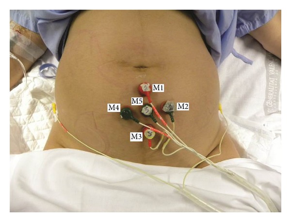
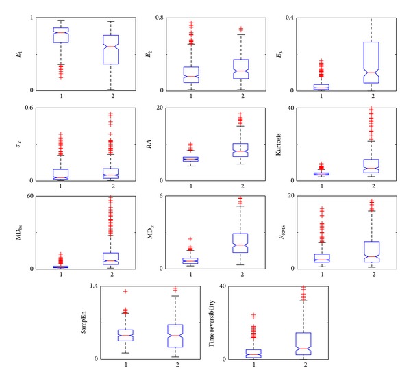
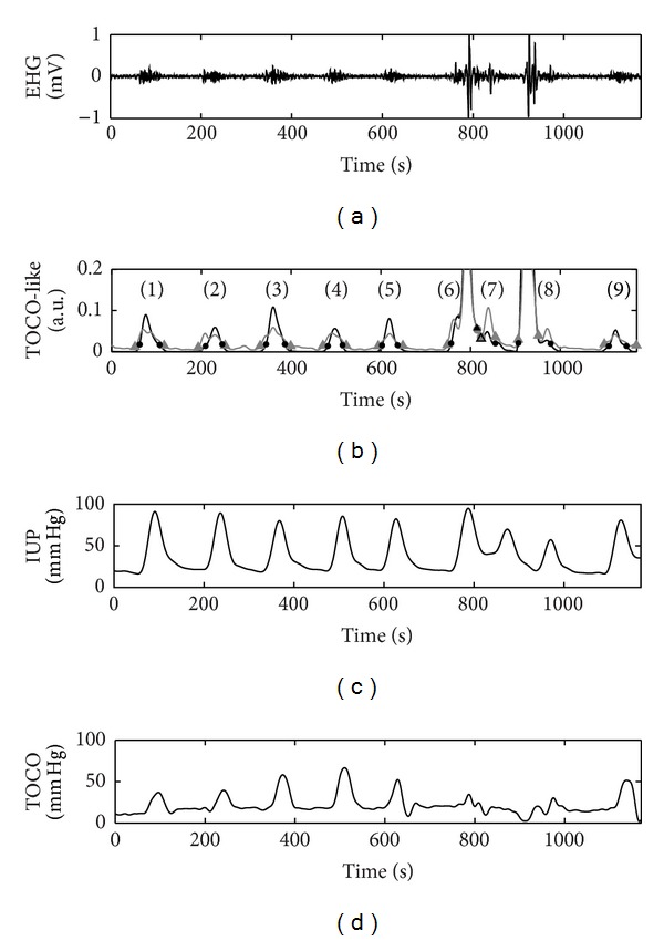
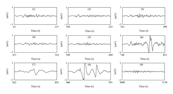
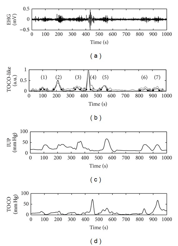
Electrohysterography (EHG) is a noninvasive technique for monitoring uterine electrical activity. However, the presence of artifacts in the EHG signal may give rise to erroneous interpretations and make it difficult to extract useful information from these recordings. The aim of this work was to develop an automatic system of segmenting EHG recordings that distinguishes between uterine contractions and artifacts. Firstly, the segmentation is performed using an algorithm that generates the TOCO-like signal derived from the EHG and detects windows with significant changes in amplitude. After that, these segments are classified in two groups: artifacted and nonartifacted signals. To develop a classifier, a total of eleven spectral, temporal, and nonlinear features were calculated from EHG signal windows from 12 women in the first stage of labor that had previously been classified by experts. The combination of characteristics that led to the highest degree of accuracy in detecting artifacts was then determined. The results showed that it is possible to obtain automatic detection of motion artifacts in segmented EHG recordings with a precision of 92.2% using only seven features. The proposed algorithm and classifier together compose a useful tool for analyzing EHG signals and would help to promote clinical applications of this technique.
Figures
Similar articles
-
Automated electrohysterographic detection of uterine contractions for monitoring of pregnancy: feasibility and prospects.BMC Pregnancy Childbirth. 2018 May 8;18(1):136. doi: 10.1186/s12884-018-1778-1. BMC Pregnancy Childbirth. 2018. PMID: 29739438 Free PMC article.
-
Automatic semantic segmentation of EHG recordings by deep learning: An approach to a screening tool for use in clinical practice.Comput Methods Programs Biomed. 2024 Sep;254:108317. doi: 10.1016/j.cmpb.2024.108317. Epub 2024 Jul 5. Comput Methods Programs Biomed. 2024. PMID: 38996804
-
Multi-channel electrohysterography enabled uterine contraction characterization and its effect in delivery assessment.Comput Biol Med. 2023 Dec;167:107697. doi: 10.1016/j.compbiomed.2023.107697. Epub 2023 Nov 8. Comput Biol Med. 2023. PMID: 37976821
-
Accuracy of frequency-related parameters of the electrohysterogram for predicting preterm delivery: a review of the literature.Obstet Gynecol Surv. 2009 Aug;64(8):529-41. doi: 10.1097/OGX.0b013e3181a8c6b1. Obstet Gynecol Surv. 2009. PMID: 19624864 Review.
-
Clinical assessment of uterine contractions.Int J Gynaecol Obstet. 2017 Nov;139(2):137-142. doi: 10.1002/ijgo.12270. Epub 2017 Aug 11. Int J Gynaecol Obstet. 2017. PMID: 28727889 Review.
Cited by
-
On the prediction of premature births in Hispanic labour patients using uterine contractions, heart beat signals and prediction machines.Healthc Technol Lett. 2023 Apr 8;10(1-2):11-22. doi: 10.1049/htl2.12044. eCollection 2023 Feb-Apr. Healthc Technol Lett. 2023. PMID: 37077881 Free PMC article.
-
Uterine electromyography for discrimination of labor imminence in women with threatened preterm labor under tocolytic treatment.Med Biol Eng Comput. 2019 Feb;57(2):401-411. doi: 10.1007/s11517-018-1888-y. Epub 2018 Aug 29. Med Biol Eng Comput. 2019. PMID: 30159659
-
Assessment of Features between Multichannel Electrohysterogram for Differentiation of Labors.Sensors (Basel). 2022 Apr 27;22(9):3352. doi: 10.3390/s22093352. Sensors (Basel). 2022. PMID: 35591042 Free PMC article.
-
A Comparative Study of Vaginal Labor and Caesarean Section Postpartum Uterine Myoelectrical Activity.Sensors (Basel). 2020 May 26;20(11):3023. doi: 10.3390/s20113023. Sensors (Basel). 2020. PMID: 32466584 Free PMC article.
-
Enhancing uterine contraction detection through novel EHG signal processing: a pilot study leveraging the relationship between slow and fast wave components to improve signal quality and noise resilience.Front Physiol. 2025 May 27;16:1568919. doi: 10.3389/fphys.2025.1568919. eCollection 2025. Front Physiol. 2025. PMID: 40496244 Free PMC article.
References
-
- Wilmink FA, Wilms FF, Heydanus R, Mol BWJ, Papatsonis DNM. Fetal complications after placement of an intrauterine pressure catheter: a report of two cases and review of the literature. Journal of Maternal-Fetal and Neonatal Medicine. 2008;21(12):880–883. - PubMed
-
- Vinken MP, Rabotti C, Mischi M, Oei SG. Accuracy of frequency-related parameters of the electrohysterogram for predicting preterm delivery: a review of the literature. Obstetrical and Gynecological Survey. 2009;64(8):529–541. - PubMed
-
- Schlembach D, Maner WL, Garfield RE, Maul H. Monitoring the progress of pregnancy and labor using electromyography. European Journal of Obstetrics Gynecology and Reproductive Biology. 2009;144(supplement 1):S33–S39. - PubMed
-
- Miles AM, Monga M, Richeson KS. Correlation of external and internal monitoring of uterine activity in a cohort of term patients. American Journal of Perinatology. 2001;18(3):137–140. - PubMed
-
- Devedeux D, Marque C, Mansour S, Germain G, Duchene J. Uterine electromyography: a critical review. American Journal of Obstetrics and Gynecology. 1993;169(6):1636–1653. - PubMed
Publication types
MeSH terms
LinkOut - more resources
Full Text Sources
Other Literature Sources
