. 2014 Feb 11;9(2):e87684.
doi: 10.1371/journal.pone.0087684. eCollection 2014.

BRK targets Dok1 for ubiquitin-mediated proteasomal degradation to promote cell proliferation and migration

Affiliations

BRK targets Dok1 for ubiquitin-mediated proteasomal degradation to promote cell proliferation and migration

Sayem Miah et al. PLoS One. .

Erratum in

  • PLoS One. 2014;9(5):e98814

Abstract

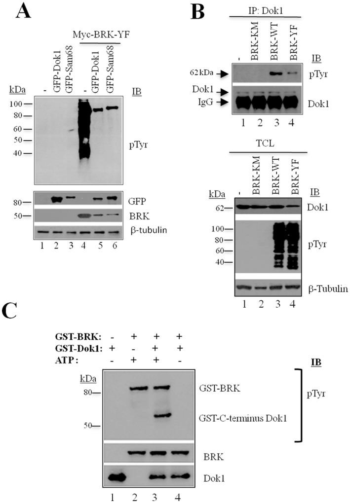
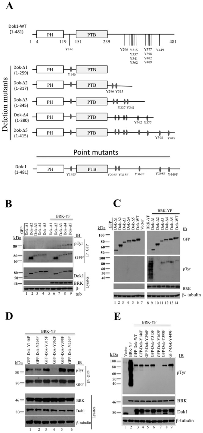
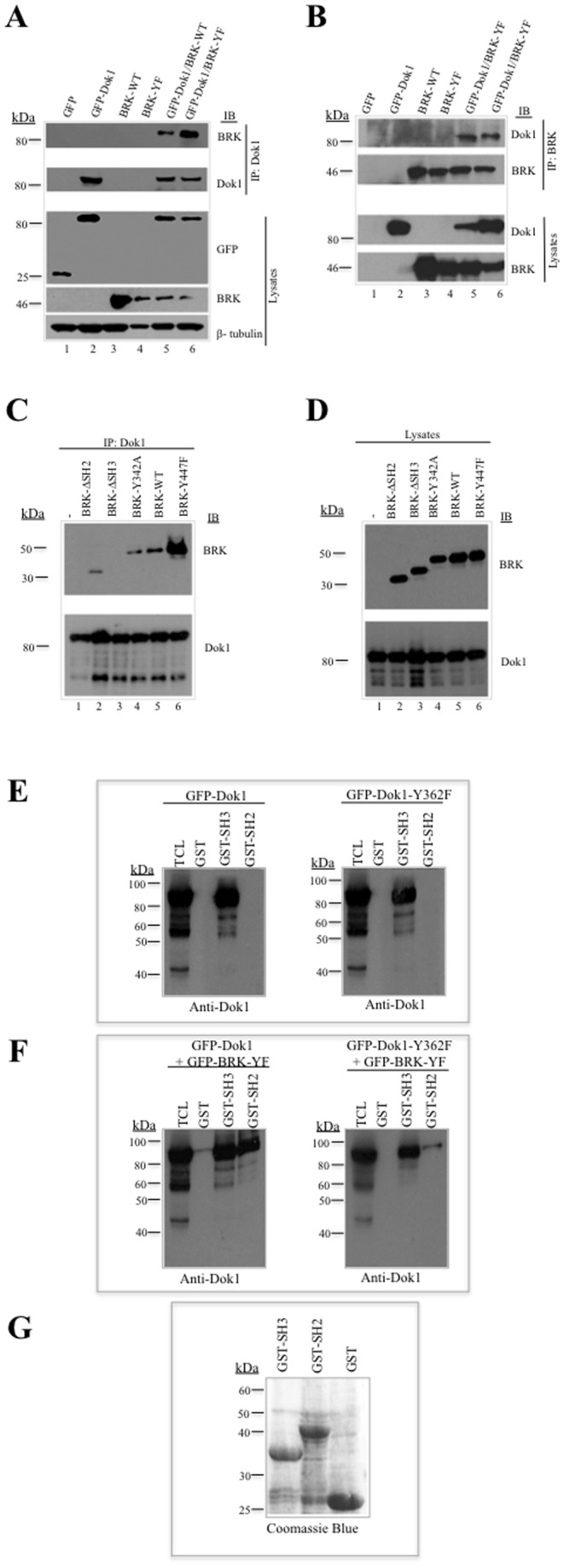
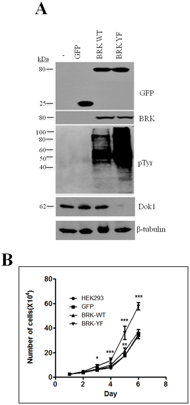
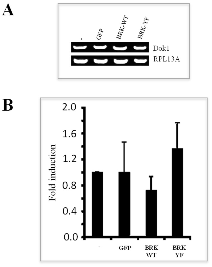
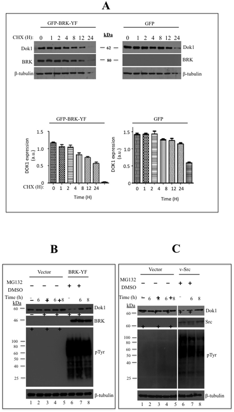
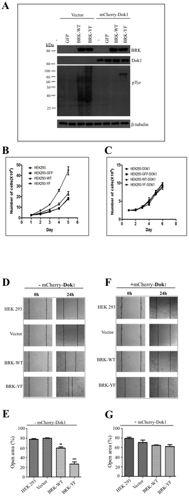
Breast tumor kinase (BRK), also known as protein tyrosine kinase 6 (PTK6), is a non-receptor tyrosine kinase overexpressed in more that 60% of human breast carcinomas. The overexpression of BRK has been shown to sensitize mammary epithelial cells to mitogenic signaling and to promote cell proliferation and tumor formation. The molecular mechanisms of BRK have been unveiled by the identification and characterization of BRK target proteins. Downstream of tyrosine kinases 1 or Dok1 is a scaffolding protein and a substrate of several tyrosine kinases. Herein we show that BRK interacts with and phosphorylates Dok1 specifically on Y362. We demonstrate that this phosphorylation by BRK significantly downregulates Dok1 in a ubiquitin-proteasome-mediated mechanism. Together, these results suggest a novel mechanism of action of BRK in the promotion of tumor formation, which involves the targeting of tumor suppressor Dok1 for degradation through the ubiquitin proteasomal pathway.


Conflict of interest statement

Competing Interests: The authors have declared that no competing interests exist.

Figures

Figure 1
Figure 1. Dok1 is a direct substrate of BRK.
(A) HEK 293 cells were transiently transfected with empty control vector (−) or GFP-Dok1, GFP-Sam68, Myc-BRK or co-transfected with Myc-BRK+GFP-Sam68 and Myc-BRK+GFP-Dok1. Tyrosine phophorylation of cellular proteins were detected in total cell lysates by immunoblot analysis (IB) with anti-phosphotyrosine (anti-pTyr) antibody (PY20). The blots were reprobed with anti-GFP, anti-BRK and anti-β- Tubulin antibodies as a loading control. (B) Tyrosine phosphorylated endogenous Dok1 as confirmed by anti-Dok1 immunoprecipitation (IP) followed by immunoblot analysis with anti-phosphotyrosine antibody and anti-Dok1(top panel). Immunoblot analysis of total cell lysates is showing the expression of Dok1, kinase activity of BRK-WT and BRK-YF, and β-tubulin as a loading control (bottom panel). (C) An in vitro kinase assay was performed using the active kinase, GST-BRK, and the substrate, GST-C-terminus Dok1, in the presence (+) or absence (−) of ATP. Tyrosine phosphorylation was detected using anti-phosphotyrosine antibody. The blots were reprobed with anti-BRK and anti-Dok1 antibody(bottom panel).
Figure 2
Figure 2. Constitutively active BRK phosphorylates Dok1 at Y362.
(A) Schematic diagram of Dok1 showing different deletion and point mutants. (B) The Dok1 deletion mutants and BRK-YF were co-transfected in to HEK 293 cells, the cell were then subjected to immunoprecipitation with anti-GFP antibody followed by immunobloting analysis using anti-phosphotyrosine and anti-GFP antibodies (top panel). Lower panel shows the expression of different GFP-Dok1 deletion mutants, BRK (as input) and β-tubulin as a loading control. (C) Dok1 deletion mutants were transfected either alone or with BRK-YF into HEK 293 cells, the cell lysates were then subjected to immunobloting analysis using antibodies against Dok1, phosphotyrosine, BRK and β-tubulin as loading control. (D) HEK 293 cells were co-transfected with Dok1 point mutants and BRK-YF followed by immunoprecipitation with anti-Dok1 antibody and immunoblotting analysis using anti-phosphotyrosines and anti-Dok1 antibodies. Lower panel shows the expression of BRK, GFP-Dok1 mutants (as input) and β-tubulin as a loading control. (E) HEK 293 cells were cotransfected with BRK-YF and Dok1 point mutants or transfected with BRK-YF alone. Total cell lysates were analyzed by immunoblotting analysis with antibodies against phosphotyrosines, BRK, Dok1 and β-tubulin as loading control.
Figure 3
Figure 3. BRK interacts with Dok1 through the SH3 domain in vivo and in vitro.
(A) HEK 293 cells were transfected with empty vector, Myc-BRK-WT, Myc-BRK-YF, GFP-Dok1 or co-transfected with Myc-BRK-WT/GFP-Dok1 or Myc-BRK-YF/GFP-Dok1 and subjected to immunoprecipitation with anti-Dok1 and immunoblotted with BRK and Dok1 (top 2 panels). The expression of cellular proteins was determined in total cell lysates by immunoblotting for GFP, BRK and β-tubulin as loading control. (B) BRK was immunoprecipitated with anti-BRK and subjected to immunoblotting analysis with anti-phosphotyrosine, anti-Dok1 and anti-BRK antibodies (top panels). Total cell lysates indicate the expression of BRK and Dok1 proteins. (C &D) HEK 293 cells were transfected with GFP-Dok1 alone or cotransfected with the idicated mutants of BRK and subjected to immunoprecipitation with anti-Dok1 followed by immunoblotting analysis with anti-BRK and anti-Dok1 antibodies. The cellular proteins were determined from the total cell lysates by immunoblotting analysis with anti-BRK and anti-Dok1 antibodies. (E) Overexpressed GFP-Dok1 or GFP-Dok1-Y362F in HEK 293 cell lysates from GFP-Dok1 or GFP-Dok1-Y362F expressing cells were subjected to pull-down assays with GST alone or recombinant GST-SH3 or GST-SH2 domain of BRK and immunoblotting analysis was performed with anti-Dok1 antibody. (F) GFP-Dok1/BRK-YF or GFP-Dok1-Y362F/BRK-YF cotransfected cohorts of HEK 293 cell lysates were subjected to pull-down assays with GST alone or GST-SH3 or GST-SH2 domain of BRK followed by immunoblotting with anti-Dok1 antibody. (G) Bacterially expressed GST, GST-SH3 and GST-SH2 domain of BRK proteins were detected via Coomassie blue staining.
Figure 4
Figure 4. BRK and Dok1 are differentially overexpressed in the human breast cancer cell lines.
(A) Cellular proteins were detected in total cell lysates by immunoblotting analysis with anti-Dok1 and anti-BRK antibodies. β-tubulin expression served as a loading control. (B & C) SKBR3 and BT20 cells were fractionated into the cytosolic, membrane, nuclear and cytoskeleton fractions and subjected to immunoblotting analysis for the detection of BRK and Dok1. β-tubulin and Sam68 were used as controls for the cytosolic/membrane and nuclear compartments, respectively. (D) Stable BRK knockdown was performed on parental breast cancer cell lines SKBR3 using shRNA lentiviral vector plasmids against BRK and analyzed as indicated.
Figure 5
Figure 5. Constitutively active BRK downregulates Dok1 protein expression.
(A) Immunobloting analysis of total cell lysates from HEK-293 stable cell lines is showing the expression of GFP alone' GFP-BRK-WT and GFP-BRK-YF (top panel), BRK (middle panel) and phosphorylated tyrosines (bottom panel). β-tubulin served as a loading control. (B) Immunobloting analysis of endogenous Dok1 in the stable HEK293 sublines. Expression of Dok1 was examined by immunoblotting analysis. (C) Characterization of cell proliferation in response to BRK-WT and BRK-YF. The P-values were determined for control and stably transfected cells and set at ***P≥0.0001, **P≥0.001 and *P≥0.05 for statistical significance.
Figure 6
Figure 6. Constitutively active BRK does not affect the levels of Dok1 mRNA.
(A & B) Total RNA was isolated from HEK 293 cells stably transduced with empty vector, GFP, GFP-BRK-WT and GFP-BRK-YF. Levels of Dok1 mRNA were then analyzed using RT-PCR (A) and qPCR (B). RPL13A gene was used as internal control. Error means are ± SEM of three biological repeats each having three technical repeats.
Figure 7
Figure 7. Activated BRK downregulates Dok1 by reducing its stability.
(A) HEK 293 cells or HEK 293-BRK-YF stable cell line were treated with a protein synthesis inhibitor cyclohexamide (CHX: 200 µg/ml) for the indicated time points and then the cells were lysed and analyzed by immunoblotting for Dok1, BRK and β-tubulin as a loading control. (B) HEK 293 cells were stably transduced with HEK293-BRK-YF and treated with either a proteosome inhibitor MG132 (10 µM) or the vehicle DMSO as the control, at different time points (above the plot). Cellular proteins were determined in total cell lysates by immunoblotting analysis with anti-Dok1, anti-BRK, anti-phosphotyrosine antibodies. β-tubulin was used as a loading control. (C) Empty vector or V-Src was transiently transfected into HEK293 cells and the cells treated with a proteosome inhibitor MG132 (10 µM) and vehicle control DMSO for the indicated time points. Immunoblotting analysis of total cell lysates was performed to detect Dok1, v-Src, phosphotyrosines and β-tubulin served as a loading control. (D & E) HEK 293 cells were transfected with empty control vector or BRK-YF or v-Src and treated with MG132 (10 µM) and Lactacystin (5 µM) or control vehicle for 8 hours. Then the cell lysates were subjected to immunoblot analysis with anti-Dok1 antibody. β-tubulin as a loading control. (F) HEK293-BRK-YF stable cells were transiently cotransfected with Dok1 and HA-Ubiquitin plasmids and after 12 hours the cells were treated MG132 (10 µM) for an additional 8 hours. The total cell lysates were subjected to immunoprecipitation with anti-Dok1 followed by immunoblotting analysis with anti-HA and anti-Dok1 antibodies. The inputs were analysed as indicated.
Figure 8
Figure 8. Dok1 inhibits BRK-induced cell proliferation and migration.
(A) HEK 293 stable sub-cell lines were transduced with mCherry-Dok1 using adenoviral vector. Cellular proteins were detected in total cell lysates by immunoblotting analysis with anti-BRK, anti-Dok1, and anti-phosphotyrosine antibodies. β-tubulin served as a loading control. (B & C) HEK 293 stable cells were transduced with or without mCherry-Dok1adeno-vector and were monitored for cell proliferation. (D & E) Cell migration determined by the healing of a fixed wound area induced in the different HEK 293 stable transfectant cells. The percentage of open area at 24 h is plotted. (F & G) Cell migration analysis was performed with the indicated stable cell lines expressing mCherry-Dok1 or an empty vector. The assay was based on the rate of wound closure in the scratched cells. The percentage of open area at 24 hours is plotted. The migration assay was performed in three independent experiments. Data are means ± standard errors. Statistics: and **P≥0.001 and ***P≥0.0001.

References

    1. Lee S-K, Shaw AS, Maher SE, Bothwell ALM (1993) p59fyn tyrosine kinase is an essential regulator of p56lck tyrosine kinase activity and early TcR-mediated signalling. - PubMed
    1. Mitchell PJ, Barker KT, Martindale JE, Kamalati T, Lowe PN, et al. (1994) Cloning and characterisation of cDNAs encoding a novel non-receptor tyrosine kinase, brk, expressed in human breast tumours. Oncogene 9: 2383–2390. - PubMed
    1. Barker KT, Jackson LE, Crompton MR (1997) BRK tyrosine kinase expression in a high proportion of human breast carcinomas. Oncogene 15: 799–805. - PubMed
    1. Mitchell PJ, Barker KT, Martindale JE, Kamalati T, Lowe PN, et al. (1994) Cloning and characterization of cDNAs encoding a novel non-receptor tyrosine kinase, brk, expressed in human breast tumours. Oncogene 9: 2383–2390. - PubMed
    1. Ostrander JH, Daniel AR, Lofgren K, Kleer CG, Lange CA (2007) Breast tumor kinase (protein tyrosine kinase 6) regulates heregulin-induced activation of ERK5 and p38 MAP kinases in breast cancer cells. Cancer Res 67: 4199–4209. - PubMed

Publication types

MeSH terms