Lignans from the bark of Eucommia ulmoides inhibited Ang II-stimulated extracellular matrix biosynthesis in mesangial cells
- PMID: 24524265
- PMCID: PMC3937011
- DOI: 10.1186/1749-8546-9-8
Lignans from the bark of Eucommia ulmoides inhibited Ang II-stimulated extracellular matrix biosynthesis in mesangial cells
Abstract
Background: Tree bark of Eucommia ulmoides Oliv., (commonly well-known as "Du-zhong" in China), has been used to treat hypertension, hypercholesterolemia, hyperglycemia, hepatic fibrosis and renal injury. This study aims to investigate the effects of lignans extracted from the bark of Eucommia ulmoides Oliv. on Ang II-induced proliferation and extracellular matrix biosynthesis in rat mesangial cells.
Methods: Rat mesangial cells (RMCs) were cultured in vitro and divided into six groups (control, Ang II, losartan, and low, middle and high concentration lignans groups). RMC proliferation was measured by MTT assay. RT-qPCR and western blotting were used to detect mRNA and protein expression of collagen type I (Col I), collagen type III (Col III), collagen type IV (Col IV), fibronectin and aldose reductase (AR).
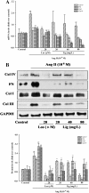
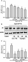
Results: Cellular proliferation induced by Ang II was significantly suppressed by Eucommia lignans of different concentrations (P = 0.034, P < 0.001, and P < 0.001). Treatment of cells with Ang II increased Col I, Col III, Col IV, and fibronectin mRNA expression, which was observed at the protein level (P < 0.001, P < 0.001, P = 0.004, and P = 0.004, respectively). The increased mRNA expression and protein levels of Col I, Col III, Col IV, and fibronectin were diminished remarkably with by treatment Eucommia lignans, and elevated AR expression stimulated by Ang II was significantly inhibited by Eucommia lignans.
Conclusions: Eucommia lignans (Du-zhong) inhibited Ang II-stimulated extracellular matrix biosynthesis in mesangial cells.
Figures


Similar articles
-
Eucommia ulmoides Oliv. (Du-Zhong) Lignans Inhibit Angiotensin II-Stimulated Proliferation by Affecting P21, P27, and Bax Expression in Rat Mesangial Cells.Evid Based Complement Alternat Med. 2015;2015:987973. doi: 10.1155/2015/987973. Epub 2015 Jun 10. Evid Based Complement Alternat Med. 2015. PMID: 26170892 Free PMC article.
-
Protective effects of Eucommia lignans against hypertensive renal injury by inhibiting expression of aldose reductase.J Ethnopharmacol. 2012 Jan 31;139(2):454-61. doi: 10.1016/j.jep.2011.11.032. Epub 2011 Nov 26. J Ethnopharmacol. 2012. PMID: 22138658
-
Eucommia ulmoides Oliv.: ethnopharmacology, phytochemistry and pharmacology of an important traditional Chinese medicine.J Ethnopharmacol. 2014;151(1):78-92. doi: 10.1016/j.jep.2013.11.023. Epub 2013 Dec 1. J Ethnopharmacol. 2014. PMID: 24296089 Review.
-
Antihypertensive Activity of Eucommia Ulmoides Oliv: Male Flower Extract in Spontaneously Hypertensive Rats.Evid Based Complement Alternat Med. 2020 Apr 30;2020:6432173. doi: 10.1155/2020/6432173. eCollection 2020. Evid Based Complement Alternat Med. 2020. PMID: 32419815 Free PMC article.
-
Research Progress of Eucommia ulmoides Oliv and Predictive Analysis of Quality Markers Based on Network Pharmacology.Curr Pharm Biotechnol. 2024;25(7):860-895. doi: 10.2174/0113892010265000230928060645. Curr Pharm Biotechnol. 2024. PMID: 38902931 Review.
Cited by
-
Efficacy and safety of Qiangli Dingxuan tablet combined with amlodipine besylate for essential hypertension: a randomized, double-blind, placebo-controlled, parallel-group, multicenter trial.Front Pharmacol. 2023 Jul 10;14:1225529. doi: 10.3389/fphar.2023.1225529. eCollection 2023. Front Pharmacol. 2023. PMID: 37492087 Free PMC article.
-
Eucommia bark (Du-Zhong) improves diabetic nephropathy without altering blood glucose in type 1-like diabetic rats.Drug Des Devel Ther. 2016 Mar 2;10:971-8. doi: 10.2147/DDDT.S98558. eCollection 2016. Drug Des Devel Ther. 2016. PMID: 27041999 Free PMC article.
-
Network Pharmacology-Based Prediction and Verification of the Potential Targets of Pinoresinol Diglucoside for OA Treatment.Evid Based Complement Alternat Med. 2022 Apr 16;2022:9733742. doi: 10.1155/2022/9733742. eCollection 2022. Evid Based Complement Alternat Med. 2022. PMID: 35469160 Free PMC article.
-
Amelioration of Bleomycin-induced Pulmonary Fibrosis of Rats by an Aldose Reductase Inhibitor, Epalrestat.Korean J Physiol Pharmacol. 2015 Sep;19(5):401-11. doi: 10.4196/kjpp.2015.19.5.401. Epub 2015 Aug 20. Korean J Physiol Pharmacol. 2015. PMID: 26330752 Free PMC article.
-
Eucommia ulmoides Oliv. (Du-Zhong) Lignans Inhibit Angiotensin II-Stimulated Proliferation by Affecting P21, P27, and Bax Expression in Rat Mesangial Cells.Evid Based Complement Alternat Med. 2015;2015:987973. doi: 10.1155/2015/987973. Epub 2015 Jun 10. Evid Based Complement Alternat Med. 2015. PMID: 26170892 Free PMC article.
References
-
- 2009 Annual data report: atlas of end-stage renal disease in the United States. [ http://www.usrds.org/2009/slides/indiv/INDEX_CKD.HTML]
LinkOut - more resources
Full Text Sources
Other Literature Sources
Research Materials
Miscellaneous

