Review
. 2014 Mar;41(1):1-24.
doi: 10.1016/j.clp.2013.11.001.

Cerebral white and gray matter injury in newborns: new insights into pathophysiology and management

Affiliations
Review

Cerebral white and gray matter injury in newborns: new insights into pathophysiology and management

Stephen A Back. Clin Perinatol. 2014 Mar.

Abstract

Increasing numbers of preterm neonates survive with motor and cognitive disabilities related to less destructive forms of cerebral injury that still result in reduced cerebral growth. White matter injury results in myelination disturbances related to aberrant responses to death of pre-myelinating oligodendrocytes (preOLs). PreOLs are rapidly regenerated but fail to mature to myelinating cells. Although immature projection neurons are more resistant to hypoxia-ischemia than preOLs, they display widespread disturbances in dendritic arbor maturation, which provides an explanation for impaired cerebral growth. Thus, large numbers of cells fail to fully mature during a critical window in development of neural circuitry. These recently recognized forms of cerebral gray and white matter dysmaturation suggest new therapeutic directions centered on reversal of the processes that promote dysmaturation.

Keywords: Cerebral; Gray matter; Management; Newborns; Pathophysiology; White matter.


Figures

Figure 1
Figure 1
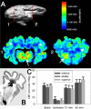
Preterm fetal cerebral blood flow (CBF) does not display gradients of flow under basal or ischemia-reperfusion conditions that is consistent with vascular border zones or endzones. (A) Quantification of regional fetal cerebral blood flow in vivo under conditions of basal flow. The top image in panel A represents a 3-D surface reconstruction of fluorescence images of a 0.65 gestation control ovine brain that indicates the frontal and parietal levels to which the lower blood flow images correspond in 1 and 2. Representative pseudocolor scale basal flow images show higher blood flow (arrows) in the pons (image 1) and sub-cortical gray matter (image 2) and lower flow (dark blue) in the periventricular white matter (arrowheads). (B) The fetal cerebral white matter was segmented into medial and lateral sections, both of which were further segmented into inferior, middle, and superior regions. No differences were found between basal CBF values in medial and lateral white matter. (C) Basal CBF values (mean ± SEM) for the entire inferior, middle and superior PVWM. No differences in CBF were seen between superior and inferior regions of cerebral white matter, which supported a lack of gradients of CBF during basal or ischemia-reperfusion conditions. Adapted from Riddle A, Luo N, Manese M, et al. Spatial heterogeneity in oligodendrocyte lineage maturation and not cerebral blood flow predicts fetal ovine periventricular white matter injury. J Neurosci. 2006;26:3045-55 and McClure M, Riddle A, Manese M, et al. Cerebral blood flow heterogeneity in preterm sheep: lack of physiological support for vascular boundary zones in fetal cerebral white matter. J Cereb Blood Flow Metab. 2008;28(5):995-1008 with permission.
Figure 2
Figure 2
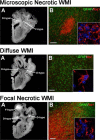
Three forms of high field MRI-defined perinatal WMI with corresponding histopathological features that were generated in the 0.65 gestation fetal sheep brain at 1 or 2 weeks after global cerebral ischemia Adapted from Riddle A, Dean J, Buser JR, et al. Histopathological correlates of magnetic resonance imaging-defined chronic perinatal white matter injury. Ann Neurol. 2011 Sep;70(3):493-507; with permission. Upper Panel. Microscopic necrotic WMI. (A) Representative appearance of a focal hypointense (F-hypo) lesion seen on a T2w image at 2 weeks after injury. Note the substantial difference in the F-hypo lesion relative to a diffuse gliotic lesion at 2 weeks, which appears more hyperintense (D-hyper). (B) A typical microscopic necrotic lesion defined by a discrete focus of immunohistochemical staining for reactive microglia and macrophages with Iba1 (red and inset) and a paucity of staining for astrocytes with glial fibrillary acidic protein (GFAP; green). Nuclei in the inset are visualized with Hoechst 33342 (blue). Bar in B, 100 μm. Middle Panel: Diffuse WMI (A) Representative appearance and distribution of diffuse hypointense (D-hypo) lesions seen on a T2w image at 1 week after injury. (B) Diffuse WMI had pronounced astrogliosis defined by immunohistochemical staining of reactive astrocytes with glial fibrillary acidic protein (GFAP; green) and a lesser population of Iba1-labeled microglia/macrophages (red) with a reactive morphology (inset). Nuclei in the inset are visualized with Hoechst 33342 (blue). Bar in B, 100 μm. Lower Panel. Focal Necrotic WMI. (A) Representative appearance from the largest focal hyperintense (F-hyper) lesion seen on a T2w image at 1 week after injury. These lesions typically localized to subcortical white matter. Note the substantial difference in the F-hyper lesion relative to the diffuse gliotic lesions, which appears much more hypointense (D-hypo). (B) A typical macroscopic necrotic lesion defined by diffuse dense staining for reactive microglia and macrophages with Iba1 (red and inset) and a paucity of GFAP-labeled astrocytes. Nuclei in the inset are visualized with Hoechst 33342 (blue). Bar in B, 100 μm.
Figure 3
Figure 3
Numerous late oligodendrocyte progenitors (preOLs) accumulate in chronic myelin-deficient perinatal white matter lesions. Lesions were generated in response to unilateral hypoxia-ischemia in the postnatal day 3 (P3) rat with the contralateral hemisphere serving as control. (A) Normal early myelination (O1-antibody; green) in control subcortical white matter (corpus callosum/external capsule) at P10 is seen with low levels of GFAP-labeled astrocytes (red) mostly concentrated over the white matter. (B) Absence of myelin in the contralateral post-ischemic lesion coincided with a diffuse glial scar that stained for GFAP-labeled astrocytes. (C) Distinctly different pathogenetic mechanisms mediate impaired myelination in necrotic lesions (PVL; upper pathway) vs. lesions with diffuse gliosis (diffuse WMI; lower pathway). Hypoxiaischemia (H-I) is illustrated as one potential trigger for WMI. More severe H-I triggers white matter necrosis (upper pathway) with pan-cellular degeneration that depletes the white matter of glia and axons. Severe necrosis results in cystic PVL, whereas milder necrosis results in microcysts. Milder H-I (lower pathway) selectively triggers early preOL death. PreOLs are rapidly regenerated from a pool of early OL progenitors that are resistant to H-I. Chronic lesions are enriched in reactive glia (astrocytes and microglia/macrophages) that generate inhibitory signals that block preOLs differentiation to mature myelinating OLs. Myelination failure in diffuse WMI thus results from preOL arrest rather than axonal degeneration. The molecular mechanisms that trigger preOL arrest are likely to be multifactorial and related to factors intrinsic and extrinsic to the preOLs. Note that the lower pathway is the dominant one in most contemporary preterm survivors, whereas the minor upper pathway reflects the declining burden of white matter necrosis that has accompanied advances in neonatal intensive care.
Figure 4
Figure 4
The preterm brain is enriched in immature neurons that do not degenerate in response to ischemia, but are highly susceptible to impaired maturation that manifests as a less mature dendritic arbor with reduced spine density. (A) A typical pyramidal neuron from the preterm cerebral cortex of a control fetal sheep. Note the paucity of processes in contrast to the highly complex dendritic arbor of a pyramidal neuron from a near-term animal (B). (C, D) In response to preterm ischemia, cortical pyramidal neurons display disrupted maturation. Note that the typical control cell (C) is more highly arborized in contrast to the response to transient cerebral ischemia that resulted in a more simplified dendritic arbor (D). The relative complexity of the cells can be appreciated from the overlay of the red concentric Scholl rings, which illustrates that the processes of the dysmature neurons intersect less frequently with the rings. The yellow, white, pink, green and blue lines represent first-, second-, third-, fourth- and fifth-order branches, respectively, from the soma. Note the overall reduction in the size and complexity of the branching pattern in D.

References

    1. Synnes AR, Anson S, Arkesteijn A, et al. School Entry Age Outcomes for Infants with Birth Weight </=800 Grams. J Pediatr. 2010 Jul 31;157(6):989–94. - PubMed
    1. Wilson-Costello D, Fridedman H, Minich N, Fanaroff A, Hack M. Improved survival rates with increased neurodevelopmental disability for extremely low birth weight infants in the 1990s. Pediatrics. 2005;115(4):997–1003. - PubMed
    1. http://www.marchofdimes.com/mission/prematurity-campaign.aspx.
    1. Stoll BJ, Hansen NI, Adams-Chapman I, et al. Neurodevelopmental and growth impairment among extremely low-birth-weight infants with neonatal infection. Jama. 2004 Nov 17;292(19):2357–65. - PubMed
    1. Peterson BS, Vohr B, Staib LH, et al. Regional brain volume abnormalities and long-term cognitive outcome in preterm infants. JAMA. 2000 Oct 18;284(15):1939–47. - PubMed