Review
. 2014 Mar;14(2):225-44.
doi: 10.1586/14737159.2014.888313. Epub 2014 Feb 13.

Advances and challenges in biosensor-based diagnosis of infectious diseases

Affiliations
Review

Advances and challenges in biosensor-based diagnosis of infectious diseases

Mandy L Y Sin et al. Expert Rev Mol Diagn. 2014 Mar.

Abstract

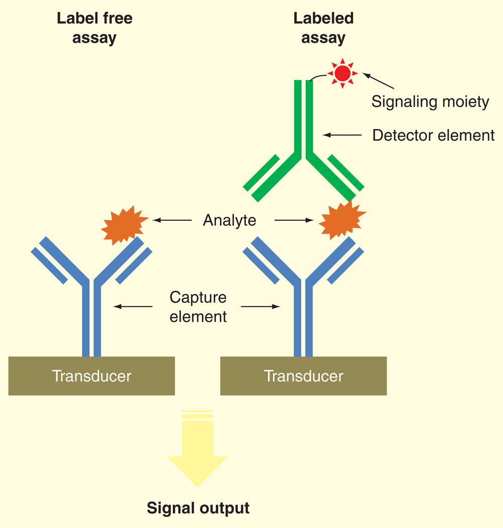
Rapid diagnosis of infectious diseases and timely initiation of appropriate treatment are critical determinants that promote optimal clinical outcomes and general public health. Conventional in vitro diagnostics for infectious diseases are time-consuming and require centralized laboratories, experienced personnel and bulky equipment. Recent advances in biosensor technologies have potential to deliver point-of-care diagnostics that match or surpass conventional standards in regards to time, accuracy and cost. Broadly classified as either label-free or labeled, modern biosensors exploit micro- and nanofabrication technologies and diverse sensing strategies including optical, electrical and mechanical transducers. Despite clinical need, translation of biosensors from research laboratories to clinical applications has remained limited to a few notable examples, such as the glucose sensor. Challenges to be overcome include sample preparation, matrix effects and system integration. We review the advances of biosensors for infectious disease diagnostics and discuss the critical challenges that need to be overcome in order to implement integrated diagnostic biosensors in real world settings.


Conflict of interest statement

Financial & competing interests disclosure

The authors have no other relevant affiliations or financial involvement with any organization or entity with a financial interest in or financial conflict with the subject matter or materials discussed in the manuscript apart from those disclosed.

No writing assistance was utilized in the production of this manuscript.

Figures

Figure 1
Figure 1
Schematic representation of label-free and labeled assays to biosensing using antibodies.
Figure 2
Figure 2. Integrated microfluidic system for multiplexed detection of HIV and syphilis
(A) Photograph of microfluidic chip. (B) Cross-section of microchannels. Scale bar, 500 µm. (C) The design of channel meanders. Scale bar, 1 mm. (D) Schematic diagram of passive fluid delivery of preloaded reagents over four detection zones based on vacuum generated by a disposable syringe. (E) Illustration of reactions at different detection steps. Signal amplification was achieved by the reduction of silver ions on gold nanoparticle-conjugated antibodies. Signals can be read quantitatively with low-cost optics or qualitatively by eyes. (F) Real-time monitoring of absorbance signals at the detection zones. Adapted with permission from [63] © Macmillan Publishers Ltd. (2011).
Figure 3
Figure 3. Integrated blood barcode chip for multiplexed detection of protein
(A) Schematic of plasma separation based on Zweifach-Fung effect from a finger prick of blood. Plasma separation channels are integrated with multiple DNA-encoded antibody barcode arrays for protein detection. (B) A, B, C represent different DNA codes. (1) is the DNA-antibody conjugate, (2) is plasma protein, (3) is biotin-labeled detection antibody, (4) is streptavidin-Cy5 fluorescence probe and (5) is complementary DNA-Cy3 reference probe. The inset is a barcode of protein biomarkers with the signal measured by fluorescence detection. The green bar denotes as an alignment marker. RBC: Red blood cells; WBC: White blood cells. Adapted with permission from [82] © Macmillan Publishers Ltd. (2008).
Figure 4
Figure 4. Integrated lab-on-a-disc platform for detection of hepatitis B virus
(A) Schematic of the disc showing the microfluidic layout and function of different compartments. The number indicates the order of operation. (B–G) Illustration of the reactions on the disc. Adapted with permission from [84] © The Royal Society of Chemistry (2009).
Figure 5
Figure 5. Demonstration of fluid manipulation with food color dyes in an integrated electrode platform for detection of bacterial 16S rRNA
(A & B) Electrolytic pumping of two color food dyes into the mixing and sensing chamber in the center. (C & D) Electrokinetic mixing of the color food dyes on top of the electrochemical sensing electrode. (E–H) Electrolytic pumping of washing buffer into the sensing chamber and delivered to the waste reservoirs. (I) Photograph of the universal electrode array for implementing the electrochemical assay for bacterial 16S rRNA. Reprinted with permission from [112] © IEEE (2013).

References

    1. Franca RF, Da Silva CC, De Paula SO. Recent advances in molecular medicine techniques for the diagnosis, prevention, and control of infectious diseases. Eur J Clin Microbiol Infect Dis. 2013;32(6):723–728. - PMC - PubMed
    1. Fauci AS, Morens DM. 200 NEJM ANNIVERSARY ARTICLE the perpetual challenge of infectious diseases. N Engl J Med. 2012;366(5):454–461. - PubMed
    1. Yager P, Domingo GJ, Gerdes J. Point-of-care diagnostics for global health. Annu Rev Biomed Eng. 2008;10:107–144. • Overview of point-of-care diagnostic technologies for global health issues

    1. Mabey D, Peeling RW, Ustianowski A, Perkins MD. Diagnostics for the developing world. Nat Rev Microbiol. 2004;2(3):231–240. - PubMed
    1. Luong JH, Male KB, Glennon JD. Biosensor technology: technology push versus market pull. Biotechnol Adv. 2008;26(5):492–500. - PubMed

Publication types