

Contributions of human tissue analysis to understanding the mechanisms of loosening and osteolysis in total hip replacement
- PMID: 24525037
- PMCID: PMC4389682
- DOI: 10.1016/j.actbio.2014.02.003
Contributions of human tissue analysis to understanding the mechanisms of loosening and osteolysis in total hip replacement
Abstract
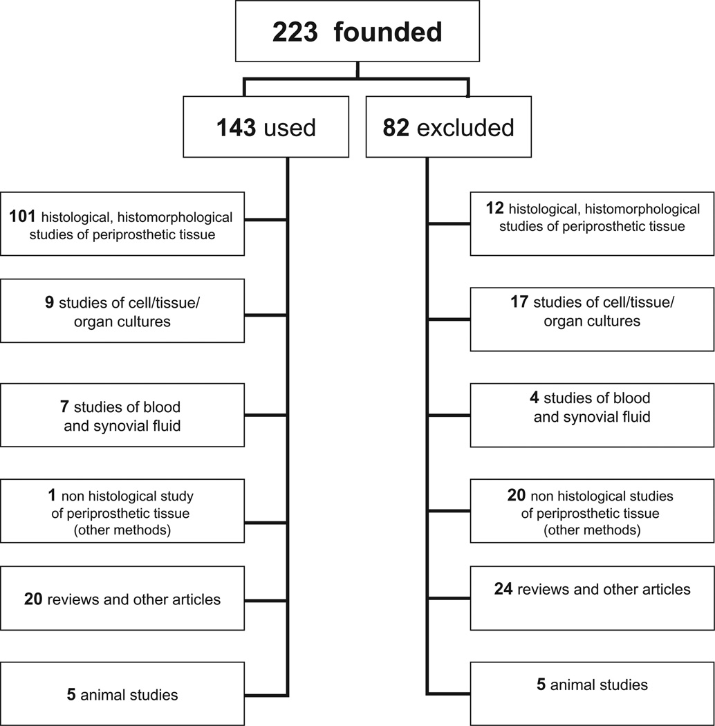
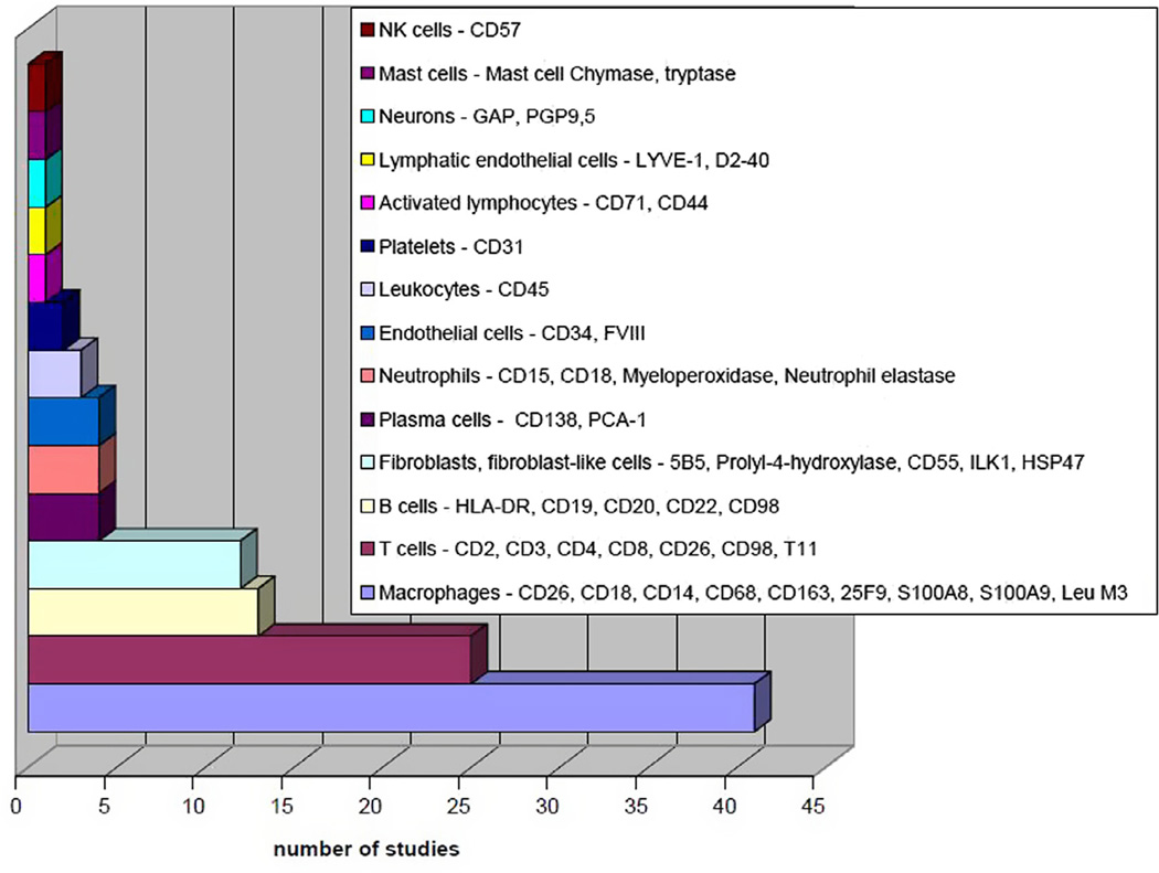
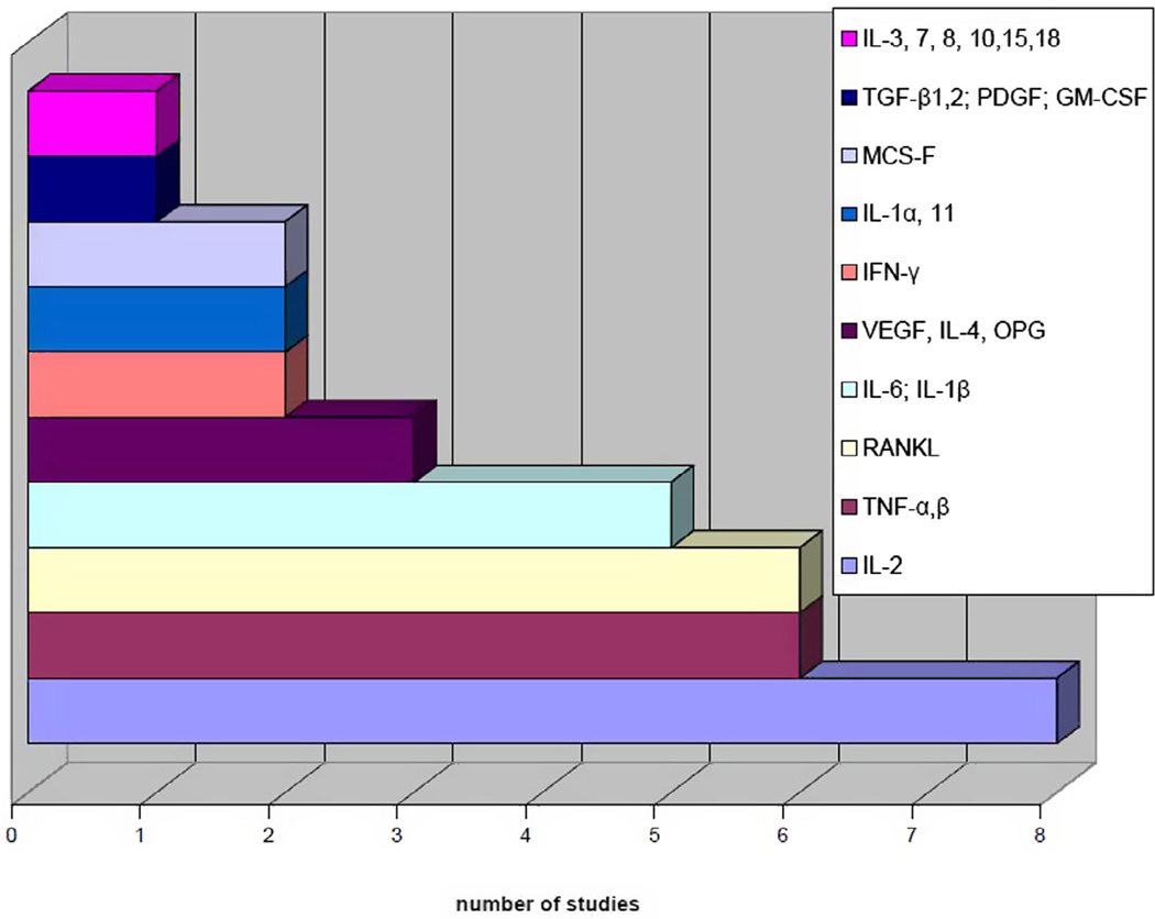
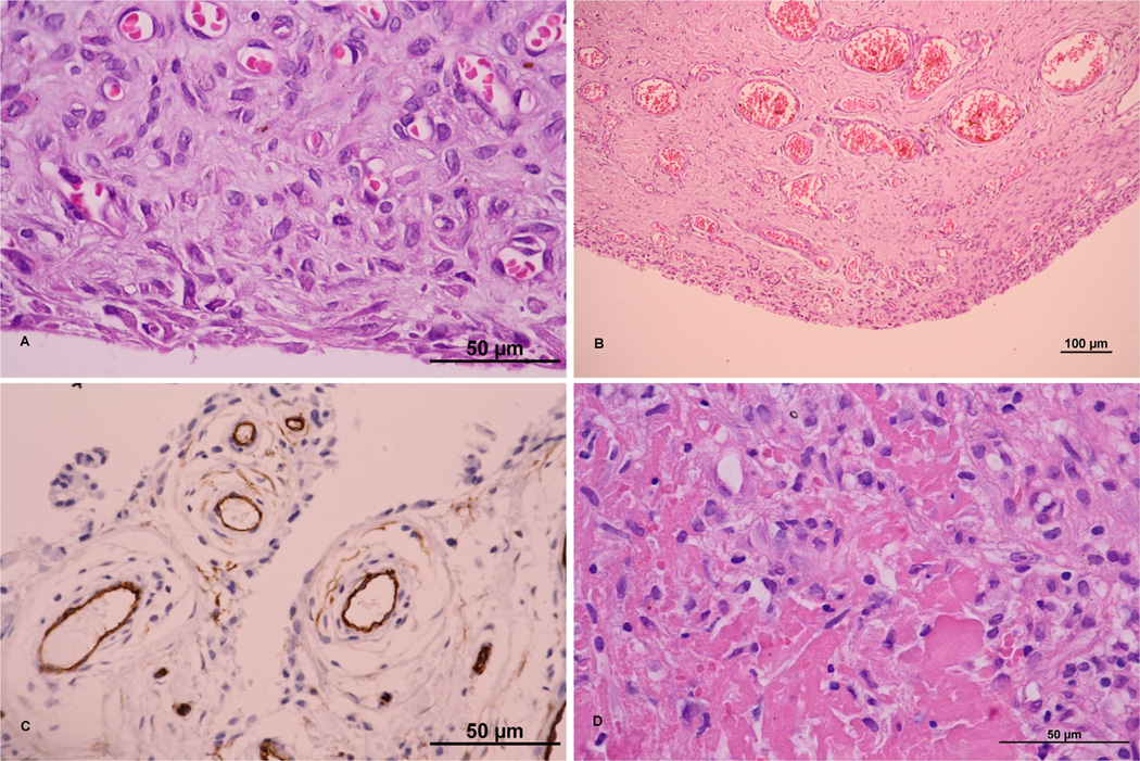
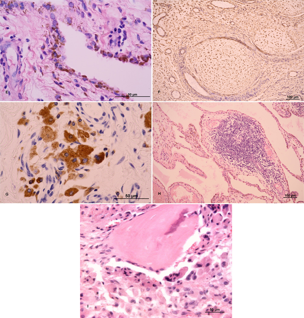
Aseptic loosening and osteolysis are the most frequent late complications of total hip arthroplasty (THA) leading to revision of the prosthesis. This review aims to demonstrate how histopathological studies contribute to our understanding of the mechanisms of aseptic loosening/osteolysis development. Only studies analysing periprosthetic tissues retrieved from failed implants in humans were included. Data from 101 studies (5532 patients with failure of THA implants) published in English or German between 1974 and 2013 were included. "Control" samples were reported in 45 of the 101 studies. The most frequently examined tissues were the bone-implant interface membrane and pseudosynovial tissues. Histopathological studies contribute importantly to determination of key cell populations underlying the biological mechanisms of aseptic loosening and osteolysis. The studies demonstrated the key molecules of the host response at the protein level (chemokines, cytokines, nitric oxide metabolites, metalloproteinases). However, these studies also have important limitations. Tissues harvested at revision surgery reflect specifically end-stage failure and may not adequately reveal the evolution of pathophysiological events that lead to prosthetic loosening and osteolysis. One possible solution is to examine tissues harvested from stable total hip arthroplasties that have been revised at various time periods due to dislocation or periprosthetic fracture in multicenter studies.
Keywords: Aseptic loosening; Immunostaining; Osteolysis; Tissue analysis; Total hip.
Copyright © 2014 Acta Materialia Inc. Published by Elsevier Ltd. All rights reserved.
Conflict of interest statement
The work has not been supported by commercial sources and we are not aware of any potential conflict of interests.
Figures
References
-
- Kurtz S, Ong K, Lau E, Mowat F, Halpern M. Projections of primary and revision hip and knee arthroplasty in the United States from 2005 to 2030. J Bone Joint Surg Am Vol. 2007;89:780–785. - PubMed
-
- Vanhegan IS, Malik AK, Jayakumar P, Ul Islam S, Haddad FS. A financial analysis of revision hip arthroplasty: the economic burden in relation to the national tariff. J Bone Joint Surg Am Br Vol. 2012;94:619–623. - PubMed
-
- Patil S, Garbuz DS, Greidanus NV, Masri BA, Duncan CP. Quality of life outcomes in revision vs primary total hip arthroplasty: a prospective cohort study. J Arthroplasty. 2008;23:550–553. - PubMed
-
- Inacio MC, Ake CF, Paxton EW, Khatod M, Wang C, Gross TP, et al. Sex and risk of hip implant failure: assessing total hip arthroplasty outcomes in the United States. JAMA Intern Med. 2013;173:435–441. - PubMed
-
- Willert HG, Ludwig J, Semlitsch M. Reaction of bone to methacrylate after hip arthroplasty: a long-term gross, light microscopic, and scanning electron microscopic study. J Bone Joint Surg Am Vol. 1974;56:1368–1382. - PubMed
Publication types
MeSH terms
Grants and funding
LinkOut - more resources
Full Text Sources
Other Literature Sources
Medical
