Review
. 2014 Apr:5:34-41.
doi: 10.1016/j.coviro.2014.01.009. Epub 2014 Feb 11.

Relating structure to evolution in class II viral membrane fusion proteins

Affiliations
Review

Relating structure to evolution in class II viral membrane fusion proteins

Yorgo Modis. Curr Opin Virol. 2014 Apr.

Abstract

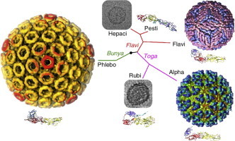
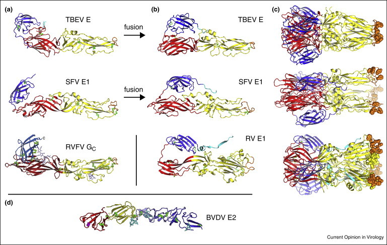
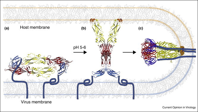
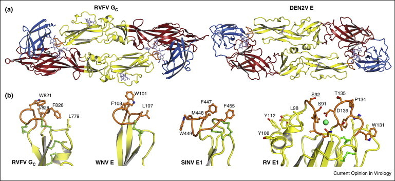
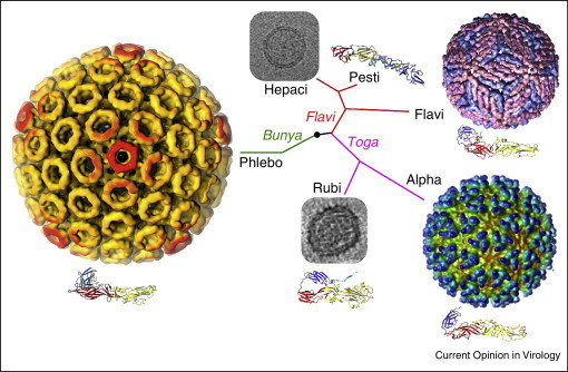
Enveloped viruses must fuse their lipid membrane to a cellular membrane to deliver the viral genome into the cytoplasm for replication. Viral envelope proteins catalyze this critical membrane fusion event. They fall into at least three distinct structural classes. Class II fusion proteins have a conserved three-domain architecture and are found in many important viral pathogens. Until 2013, class II proteins had only been found in flaviviruses and alphaviruses. However, in 2013 a class II fusion protein was discovered in the unrelated phlebovirus genus, and two unexpectedly divergent envelope proteins were identified in families that also contain prototypical class II proteins. The structural relationships of newly identified class II proteins, reviewed herein, shift the paradigm for how these proteins evolved.


Figures

None
Graphical abstract
Figure 1
Figure 1
Representative class II membrane fusion glycoproteins in their prefusion and postfusion conformations. (a) The class II fold consists of three domains. A β-sandwich domain (red) organizes the structure; an elongated domain (yellow) bears a hydrophobic “fusion loop” (orange) at its tip, which serves as an anchor in the target cellular membrane; an Ig-like domain (blue) contains the structural determinants of cellular tropism and virulence, as well as most neutralizing antibody epitopes. The following viral fusion proteins are shown in their prefusion conformation: E from the flavivirus tick-borne encephalitis virus (TBEV) [11]; E1 from the alphavirus Semliki Forest virus (SFV) [19]; Gc from Rift Valley fever virus (RVFV), a phlebovirus from the Bunyaviridae family [5••]. (b) Class II proteins in their postfusion conformation. Shown here are TBEV E [42], SFV E1 [43] and E1 from rubella virus (RV) [6••]. Class II proteins are trimeric in the postfusion conformation, (c). (d) Envelope protein E2 from bovine viral diarrhea virus (BVDV) has a novel fold despite being in the Flaviviridae family (genus pestivirus) [9••, 10••].
Figure 2
Figure 2
Membrane fusion by class II envelope proteins. (a) The protein forms dimers in the outer protein shell of the virion. The “stem-anchor” (cyan) tethers the protein to the viral membrane. Gc from Rift Valley fever virus (RVFV) is shown as an example [5••]. (b) The protein responds to the reduced pH of an endosomal compartment with a hinge motion that exposes the hydrophobic fusion loop (orange). The fusion loop inserts into the cell membrane. A crystal structure of RVFV Gc proposed to correspond to this “prehairpin” intermediate is shown [5••]. (c) The protein then folds back on itself, directing the fusion loop toward the transmembrane anchor. The refolding energy bends the apposed membranes. Creation of additional trimer contacts between the stem-anchor and the ectodomain leads to fusion of the viral and cellular membranes. The postfusion conformation of dengue type 2 virus is shown [41].
Figure 3
Figure 3
Conserved structural features in class II fusion proteins. (a) Gc from Rift Valley fever virus (RVFV) crystallizes in a dimeric head-to-tail configuration [5••]. The Gc dimers are strikingly similar to the flavivirus E dimers (dengue type 2 E shown here [12]). E dimers are the building block of the icosahedral outer protein shell in flaviviruses [26]. (b) The fusion loop serves as the anchor in the target cellular membrane during the fusion reaction (see Figure 2). The structure of the fusion loop is highly conserved in class II fusion proteins. Shown here are the fusion loops of, from left to right, RVFV Gc [5••], Sindbis virus (SINV) E1 [20], West Nile virus (WNV) E [13], and rubella virus (RV) E1 [6••].
Figure 4
Figure 4
Structural relationships of viruses that contain class II fusion proteins. The class II fold is highly conserved in flaviviruses, alphaviruses and phleboviruses, even though these viruses differ in their genomic organization, coding strategies and outer protein shell assemblies. These three genera have in common that they have lifecycles that alternate between vertebrate and arthropod hosts. Rubella virus (RV) E1 has the most divergent class II fold even though rubella belongs to the same family as alphaviruses (Togaviridae). Glycoprotein E2 from the pestivirus bovine viral diarrhea virus has a novel fold even though pestiviruses belong to the same family as flaviviruses (Flaviviridae) [9••, 10••]. Rubella virus and pestiviruses, and their close relatives the hepaciviruses, have in common that they infect strictly vertebrate hosts, and also that they do not form rigid icosahedral outer protein shells. Thus, structural conservation in viral fusion proteins does not correlate with overall phylogenetic relatedness. The virus particles shown here are, clockwise from top right, dengue virus, Semliki Forest virus, RV, Rift Valley fever virus and hepatitis C virus (HCV). The electron micrographs of RV [55] and HCV [61] are not drawn to scale with the particles in color. The phylogenetic tree is based on qualitative structural and genetic relationships between envelope proteins and is not based on a quantitative phylogenetic analysis.

References

    1. Skehel J.J., Wiley D.C. Receptor binding and membrane fusion in virus entry: the influenza hemagglutinin. Annu Rev Biochem. 2000;69:531–569. - PubMed
    1. Lamb R.A., Jardetzky T.S. Structural basis of viral invasion: lessons from paramyxovirus F. Curr Opin Struct Biol. 2007;17:427–436. - PMC - PubMed
    1. Schibli D.J., Weissenhorn W. Class I and class II viral fusion protein structures reveal similar principles in membrane fusion. Mol Membr Biol. 2004;21:361–371. - PubMed
    1. Kielian M., Rey F.A. Virus membrane-fusion proteins: more than one way to make a hairpin. Nat Rev Microbiol. 2006;4:67–76. - PMC - PubMed
    1. Dessau M., Modis Y. Crystal structure of glycoprotein C from Rift Valley fever virus. Proc Natl Acad Sci U S A. 2013;110:1696–1701. - PMC - PubMed
    2. This study showed that the Gc envelope protein from Rift Valley fever virus (from the Bunyaviridae family) has a class II fold with striking resemblances to that of E from dengue and other flaviviruses, including a propensity to form head-to-tail dimers with a hydrophobic membrane anchor, or fusion loop buried at the dimer interface. RVFV Gc was the first class II protein identified in a virus family otherwise unrelated to flaviviruses and alphaviruses, suggesting that class II proteins may have been transferred as independent modules during evolution from a host or another virus.

Publication types

MeSH terms

Substances

LinkOut - more resources